Fig. 2 (shortened, full legend in paper): Structurally characterized M. oryzae effectors in complex with the HMA or HMA-like domains of host target proteins and ID-NLRs. Crystal structures of (A) AVR-PikF with the HMA-like domain of OsHIPP19 from rice (7B1I) (Maidment et al., 2021); (B) APikL2A with the HMA-like domain of sHMA25 from foxtail millet (7NLJ) (Bentham et al., 2021); (C) Pwl2 with the HMA domain of OsHIPP43 from rice (8R7D) (Zdrzałek et al., 2024); (D) AVR-PikD with the HMA-like ID of Pikp-1 from rice (5A6W) (Maqbool et al., 2015); (E) AVR-Pia with the HMA-like ID of Pikp-1 (6Q76) (Varden et al., 2019); (F) AVR1-CO39 with the HMA-like ID of RGA5 from rice (5ZNG) (Guo et al., 2018). Complexes are displayed such that the HMA/HMA-like domains are in equivalent orientations. Domains from HPP/HIPP host targets are coloured light green. Domains from ID-NLRs are coloured dark green.
⚙️🦠 REVIEW 🦠⚙️
Turley & Faulkner explore the function of plant heavy metal-associated domain-containing proteins and speculate about their functions at plasmodesmata by drawing from plant–pathogen interaction studies.
🔗 doi.org/10.1093/jxb/...
#PlantScience 🧪 Christine Faulkner
03.02.2026 10:29 — 👍 10 🔁 7 💬 0 📌 0

Fig. 1.Dynamic regulation and role of plasmodesmata (PD) in viral RNA and siRNA movement. PD regulate the symplasmic transport of molecules between cells. Viruses encode movement proteins (MPs) that are expressed in cells at the viral infection front to facilitate viral movement through PD. Tobamovirus MPs interfere with pattern-triggered immunity (PTI), a defense mechanism activated by plant receptors that recognize viral double-stranded RNA (dsRNA) and cause PD closure by callose deposition. The open state of the PD at the viral infection front, which allows the virus to spread, also allows the movement of signaling molecules, including viral and host-derived small interfering RNAs (siRNAs). As they move ahead of the infection, these siRNAs play a critical role in systemic defense signaling and the regulation of disease outcomes. MIR, miRNA-encoding gene; PTGS, post-transcriptional gene silencing; TGS, transcriptional gene silencing.
🧬🦠 REVIEW 🧬🦠
In this review, Elvira-González et al. describe how virus-induced small RNA synthesis and small RNA movement through plasmodesmata and phloem determine the outcome of viral infection in terms of disease and tolerance.
🔗 doi.org/10.1093/jxb/...
#PlantScience 🧪
02.02.2026 17:57 — 👍 13 🔁 5 💬 2 📌 0
@laueg.bsky.social Todd Blevins
02.02.2026 17:57 — 👍 0 🔁 0 💬 0 📌 0
Fig. 1.Dynamic regulation and role of plasmodesmata (PD) in viral RNA and siRNA movement. PD regulate the symplasmic transport of molecules between cells. Viruses encode movement proteins (MPs) that are expressed in cells at the viral infection front to facilitate viral movement through PD. Tobamovirus MPs interfere with pattern-triggered immunity (PTI), a defense mechanism activated by plant receptors that recognize viral double-stranded RNA (dsRNA) and cause PD closure by callose deposition. The open state of the PD at the viral infection front, which allows the virus to spread, also allows the movement of signaling molecules, including viral and host-derived small interfering RNAs (siRNAs). As they move ahead of the infection, these siRNAs play a critical role in systemic defense signaling and the regulation of disease outcomes. MIR, miRNA-encoding gene; PTGS, post-transcriptional gene silencing; TGS, transcriptional gene silencing.
🧬🦠 REVIEW 🧬🦠
In this review, Elvira-González et al. describe how virus-induced small RNA synthesis and small RNA movement through plasmodesmata and phloem determine the outcome of viral infection in terms of disease and tolerance.
🔗 doi.org/10.1093/jxb/...
#PlantScience 🧪
02.02.2026 17:57 — 👍 13 🔁 5 💬 2 📌 0
Tina Schreier
01.02.2026 10:10 — 👍 0 🔁 0 💬 0 📌 0

Fig. 2 (shortened, full legend in paper): Plasmodesmata at different cell interfaces in C4 monocot leaves. (A) In contrast to C4 eudicots, C4 monocots can be one or combination of either NADP-ME, NAD-ME, and/or PCK subtype depending on the decarboxylation enzyme present in the bundle sheath (BS). Confocal micrographs (leftmost panel) were obtained using an excitation wavelength at 633 nm and dual emission wavelengths at 650–720 nm (magenta for Photosystem II) and 720–800 nm (blue for Photosystem I) to visualize chloroplasts. Note that BS cells of C4 NADP-ME and C4 PCK leaves have agranal chloroplasts (see second panel from left) thus appear pseudo-blue under confocal microscope due to reduced Photosystem II content. The cell wall (green) was visualized at an excitation wavelength of 405 nm and an emission wavelength of 420–480 nm.
🎯🌾 DARWIN REVIEW 🌾🎯
Schreier et al. list gene targets and propose genetic strategies to begin engineering enhanced plasmodesmata-mediated cell-to-cell connectivity in crops, aiming to increase yield and improve climate resilience.
🔗 doi.org/10.1093/jxb/...
#PlantScience 🧪
01.02.2026 10:10 — 👍 26 🔁 14 💬 2 📌 0

Fig. 4.PD and PD-associated proteins involved in cell-to-cell trafficking. (A) PD have a diverse proteome, which include PD-localized proteins and EPCS proteins, shown in magenta. PD-localized proteins and EPCS proteins can facilitate viral MP movement from cell to cell. Some PD-localized proteins are located at the PM, shown in yellow, while some are located in the ER, shown in blue. (B) MPTMV movement through PD is facilitated by SYTA. SYTA interacts directly with MPTTMV and, during TMV infection, more SYTA is recruited around EPCS and PD to further cell-to cell spread. Additionally, MPTMV interacts with RTNLBs. (C) GFLV is a tubule-forming virus and passes from cell to cell as a virion. These tubules cause ER displacement from PD and widen the aperture. These tubules are formed from MPGFLV which interacts with PDLPs. PDLPs do not initiate tubule formation but stabilize the tubule in the pore. Created in BioRender. Azim, M. (2025) https://BioRender.com/vu73y8v.
🔬 REVIEW 🔬
🦠➡️🌱 In this review, Alazem et al. discuss how plant viruses recruit and use host proteins to accomplish cell-to-cell movement via plasmodesmata during infection 🦠➡️🌱
🔗 doi.org/10.1093/jxb/...
#PlantScience 🧪 @danforthcenter.bsky.social
31.01.2026 09:58 — 👍 14 🔁 3 💬 1 📌 0

Upcoming and open special issues
The special issues listed in the table below are currently open for submissions or are due to be published soon. If you have a manuscript that you think is appr
⏱️Last couple of days left to submit!
Don't miss your opportunity to publish in @jxbotany.bsky.social's upcoming special issue looking at the challenges AI has brought to data handling, how it's has transformed predictive models, and more.
Submit by 31st January: oxford.ly/4rgsfdO
30.01.2026 12:00 — 👍 1 🔁 1 💬 0 📌 0
@hutton.ac.uk University of St Andrews
30.01.2026 08:00 — 👍 0 🔁 0 💬 0 📌 0

Fig. 1.Conceptual illustration of retention of plasmodesmata (PDs) components paired with plasma membrane (PM) and endoplasmic reticulum (ER) trafficking mechanisms. (A) PM-associated proteins traffic to the bulk PM via the Golgi body and secretory pathway. (B) Ribosomes associated with the rough ER traffic ER-associated proteins to the bulk ER. (C) The specialized composition at PDs indicated with labels and illustrated with colour gradients: the cortical ER (green) and PD–ER (navy blue), and the bulk (light blue) and specialized (purple) cell wall (CW). Magenta outlining conceptualizes the properties of PD-retained proteins clustered to the PD-specific membranes.
🔎 SPECIAL ISSUE VIEWPOINT 🔎
A variety of motifs mediate protein localization at plasmodesmata. Should they be viewed as targeting or retention signals?
Barr & Tilsner explore the motifs and mechanisms underlying plasmodesmal protein localization ⚙️
🔗 doi.org/10.1093/jxb/...
#PlantScience 🧪
30.01.2026 08:00 — 👍 12 🔁 5 💬 1 📌 0

Fluorescent proteins (mTurquoise2, mEGFP, mCitrine, mScarlet-I) move between cells via plasmodesmata in the epidermis of Nicotiana benthamiana. (Image credit: Rory Greenhalgh, Jacob O. Brunkard.)
🌱 SPECIAL ISSUE EDITORIAL 🌱
🔬 Plasmodesmata (PD) are membrane-lined channels in cell walls 🔬
In this editorial, Brunkard & Burch-Smith shift focus from "which molecules move through PD?" to "which molecules are *prevented* from moving through PD?" 🔎
🔗 doi.org/10.1093/jxb/...
#PlantScience 🧪
29.01.2026 08:00 — 👍 17 🔁 8 💬 0 📌 0

The cover of Vol 77 | Issue 3 | 2026 of the Journal of Experimental Botany, Special Issue: Plasmodesmata: Current perspectives on plant intercellular
communication and signalling. Teal coloured banners border the top and bottom of the page and in the centre is an image of multicoloured Nicotiana benthamiana cells under a microscope. Fluorescent proteins (mTurquoise2, mEGFP, mCitrine, mScarlet-I) move between cells via plasmodesmata in the epidermis of N. benthamiana. (Image credit: Rory Greenhalgh, Jacob O. Brunkard.)
📣 Check out JXB's newest Special Issue 📣
📜 Issue 3 of 2026 📜
🔬 Plasmodesmata: Current perspectives on plant intercellular communication and signalling 🔬
📘 Guest edited by Jake Brunkard & Tessa Burch-Smith
🔗 academic.oup.com/jxb...
#JXBspecialissues #PlantScience 🧪 SEBiology
28.01.2026 17:10 — 👍 18 🔁 10 💬 1 📌 0

a pink penguin with a green mohawk is holding a baby penguin with a candy cane
Alt: a pink penguin with a green mohawk is holding a baby penguin with a candy cane
Achievement unlocked:
My PhD student son and I each had a manuscript accepted on the same day. 🎉
His in Experimental Physiology @expphysiol.bsky.social
Mine in Journal of Experimental Botany @jxbotany.bsky.social
I think this calls for cake! 🍰
27.01.2026 13:49 — 👍 35 🔁 1 💬 0 📌 0
Thomas WIDIEZ
27.01.2026 12:32 — 👍 0 🔁 0 💬 0 📌 0

Fig. 2 (shortened, full legend in paper): Phenotyping of Fusarium ear rot (FER) and Fusarium seedling rot (FSR) diseases caused by F. verticillioides in the maize A188 wild type (WT) and two LOX4-overexpressing lines (OE1, OE2) at maturity. (A) Representative images of FER disease. The arrows indicate the inoculation points. (B) Quantification of FER disease severity in inoculated ears on a seven-point scale, where 1 represents no disease and 7 represents up to 100% of ear rotting. (C) Concentration of total fumonisins (B1+B2+B3) in the kernels. (D) Representative images of FSR disease on 7-day-old inoculated seedlings, and (E) quantification of disease severity in mock controls and inoculated seedlings on a five-point scale, where 1 represents no disease and 5 represents rotting of the entire kernel. (F) Quantification of disease in inoculated seedlings by F. verticillioides calmodulin copy number.
🦠🌽 RESEARCH 🌽🦠
Overexpression of LOX4 in maize enhances resistance to Fusarium verticillioides via a marked increase in the production of oxylipins together with activation of the jasmonic acid biosynthetic pathway - Ottaviani et al.
🔗 doi.org/10.1093/jxb/...
#PlantScience 🧪
27.01.2026 12:32 — 👍 11 🔁 8 💬 1 📌 0

Fig. 7.Proposed model for the role of the HY5–BBX5 module in anthocyanin biosynthesis and UV-B tolerance. Upon perceiving UV-B stress, HY5 is activated and binds to the BBX5 promoter to initiate its transcriptional expression. Subsequently, the induced BBX5 transcriptionally activates the anthocyanin biosynthesis gene ANS, thereby enhancing anthocyanins production and ultimately improving plant tolerance to UV-B radiation.
☀️🍇 RESEARCH 🍇☀️
The B-box transcription factor BBX5 is up-regulated by HY5 under UV-B radiation, which activates the anthocyanin biosynthesis gene ANS, leading to enhanced production of anthocyanins and improved UV-B resistance - Liang et al.
🔗 doi.org/10.1093/jxb/...
#PlantScience 🧪
26.01.2026 18:41 — 👍 10 🔁 3 💬 0 📌 0

Graphical Abstract
Alfalfa (Medicago sativa L.) is a key forage crop valued for its adaptability and nutritional quality, yet salinity significantly limits its productivity, particularly in arid regions. Understanding early stress responses is crucial for improving resilience. Salt stress impairs leaf growth and photosynthesis, triggering complex, time-dependent signaling. Sucrose non-fermenting kinase 1 (SnRK1), a central metabolic sensor, regulates metabolism and growth under stress. We investigated the dynamics of SnRK1, sucrose, and trehalose-6-phosphate (Tre6P) during leaf expansion in a salt-tolerant alfalfa cultivar. Plants were hydroponically grown and exposed to 200 mM NaCl. Stress induced transient declines in chloroplast performance (Fv/Fm, performance index). SnRK1 activity peaked within 1 hour post-treatment (hpt), probably initiating metabolic shifts.
🧂🌱 RESEARCH 🌱🧂
Barbieri et al. uncovered a wave-like SnRK1 activation that links early biochemical shifts to downstream metabolic changes and sugar signaling disruption, revealing key mechanisms in the salt stress response of alfalfa.
🔗 doi.org/10.1093/jxb/...
#PlantScience 🧪
26.01.2026 13:17 — 👍 11 🔁 3 💬 0 📌 0

Fig. 3.Chloroplast ultrastructure of the oslsz1 mutant and wild type (WT) plants grown in different temperature chambers. (A–L) Chloroplast ultrastructure of WT (A, B, E, F, I, J) and oslsz1 (C, D, G, H, K, L) at 22 °C (A–D), 28 °C (E–H), and 34 °C (I–L). CH, chloroplast; NU, nucleus. Scale bar=5 μm (A, C, E, G, I, K) and 0.5 μm (B, D, F, H, J, L).
🌾❄️ RESEARCH 🌾❄️
A missense mutation in a rice lycopene β-cyclase reduces carotenoid production and compromises chloroplast development at low temperatures, providing new insights into plant response to low-temperature stress - Zhang et al.
🔗 doi.org/10.1093/jxb/...
#PlantScience 🧪
24.01.2026 17:09 — 👍 4 🔁 4 💬 0 📌 0

Fig. 1.Experimental design showing the number of six-well culture plates (total of 24 plates) containing cells of all Trebouxia species (S09 C0004, S19 C0005, S12 C0006, A06 C0007, A10 C0009, A04 C0010). The cells were subjected to two light regimes: either control (60 μmol photons m−2 s−1) or high light (150 μmol photons m−2 s−1), at two time periods: 1 h and 3 d. One batch of six-well plates (n=12) were designated mainly for fluorescence measurements, which consisted of three independent biological replicates for each light treatment and period. To avoid any confounding effect from fluorescence measurements, a separate batch of six-well plates (n=12) were designated for RNA extraction and subsequently gene expression analysis. Created in BioRender. Du, R. (2025)
☀️ RESEARCH ☀️
The most common lichen photobiont genus, Trebouxia, exhibits highly species-specific responses to high light conditions, indicating the complexity of the mechanisms these organisms use to withstand stress - Poquita-Du et al.
🔗 doi.org/10.1093/jxb/...
#PlantScience 🧪
25.01.2026 11:27 — 👍 3 🔁 3 💬 0 📌 0

Fig. 7.CsDREB28 transcriptionally inhibits the expression of CsSWEET15 and AtSWEET15. (A) Schematic diagram showing potential DREB-binding sites in the promoters of CsSWEE15 and AtSWEET15. (B, C) Dual-luciferase assay and GUS staining results showed that CsDREB28 inhibited the expression of CsSWEET15 and AtSWEET15 in planta. CK, control. (D) Regulatory network model of CsDREB28 in plant growth and freezing resistance. In (B, C), data are represented as the mean ±SE with six biological replicates. Asterisks indicate significant differences (**P<0.01; Student’s t-test).
❄️🍵 RESEARCH 🍵❄️
CsDREB28 inhibits freezing resistance and plant growth in tea plants by directly transcriptionally inhibiting CsSWEET15/17 to mediate sugar transport and distribution - Peng et al.
🔗 doi.org/10.1093/jxb/...
#PlantScience 🧪
25.01.2026 11:28 — 👍 11 🔁 1 💬 0 📌 0
Fig. 1.Experimental design showing the number of six-well culture plates (total of 24 plates) containing cells of all Trebouxia species (S09 C0004, S19 C0005, S12 C0006, A06 C0007, A10 C0009, A04 C0010). The cells were subjected to two light regimes: either control (60 μmol photons m−2 s−1) or high light (150 μmol photons m−2 s−1), at two time periods: 1 h and 3 d. One batch of six-well plates (n=12) were designated mainly for fluorescence measurements, which consisted of three independent biological replicates for each light treatment and period. To avoid any confounding effect from fluorescence measurements, a separate batch of six-well plates (n=12) were designated for RNA extraction and subsequently gene expression analysis. Created in BioRender. Du, R. (2025)
☀️ RESEARCH ☀️
The most common lichen photobiont genus, Trebouxia, exhibits highly species-specific responses to high light conditions, indicating the complexity of the mechanisms these organisms use to withstand stress - Poquita-Du et al.
🔗 doi.org/10.1093/jxb/...
#PlantScience 🧪
25.01.2026 11:27 — 👍 3 🔁 3 💬 0 📌 0
Fig. 3.Chloroplast ultrastructure of the oslsz1 mutant and wild type (WT) plants grown in different temperature chambers. (A–L) Chloroplast ultrastructure of WT (A, B, E, F, I, J) and oslsz1 (C, D, G, H, K, L) at 22 °C (A–D), 28 °C (E–H), and 34 °C (I–L). CH, chloroplast; NU, nucleus. Scale bar=5 μm (A, C, E, G, I, K) and 0.5 μm (B, D, F, H, J, L).
🌾❄️ RESEARCH 🌾❄️
A missense mutation in a rice lycopene β-cyclase reduces carotenoid production and compromises chloroplast development at low temperatures, providing new insights into plant response to low-temperature stress - Zhang et al.
🔗 doi.org/10.1093/jxb/...
#PlantScience 🧪
24.01.2026 17:09 — 👍 4 🔁 4 💬 0 📌 0
![Fig. 4.Correlation-based network analysis with community detection of all common metabolites. Associations were detected by Gaussian graphical modelling [either positive (blue) or negative (red)] with the thickness of the edges indicating the strength of the correlation network. A community detected approach was used with metabolites shown in the same colour when they are assigned to the same community. (A) combined dataset of all species, (B) arabidopsis, (C) wheat, (D) plantago, (E) alchemilla, (F) strawberry, and (G) melon. 2-OG, 2-oxoglutarate; 3-PGA, 3-phosphoglycerate; ADPGlc, adenosine 5′-diphosphoglucose; Fru6P, fructose 6-phosphate; Gal1P, galactose 1-phosphate; Glc1P, glucose 1-phosphate; Glc6P, glucose 6-phosphate; Gly3P, glycerine 3-phosphate; Man6P, mannose 6-phosphate; PEP, phosphoenolpyruvate; Suc6P, sucrose 6′-phosphate; Tre6P, trehalose 6-phosphate; UDPGlc, uridine 5′-diphosphoglucose.](https://cdn.bsky.app/img/feed_thumbnail/plain/did:plc:ggz22gcwkakll7efac23g33s/bafkreif67rt3neoynspx3md4lgk7nvdtridcxtd2stwro3xvd7wsvackpa@jpeg)
Fig. 4.Correlation-based network analysis with community detection of all common metabolites. Associations were detected by Gaussian graphical modelling [either positive (blue) or negative (red)] with the thickness of the edges indicating the strength of the correlation network. A community detected approach was used with metabolites shown in the same colour when they are assigned to the same community. (A) combined dataset of all species, (B) arabidopsis, (C) wheat, (D) plantago, (E) alchemilla, (F) strawberry, and (G) melon. 2-OG, 2-oxoglutarate; 3-PGA, 3-phosphoglycerate; ADPGlc, adenosine 5′-diphosphoglucose; Fru6P, fructose 6-phosphate; Gal1P, galactose 1-phosphate; Glc1P, glucose 1-phosphate; Glc6P, glucose 6-phosphate; Gly3P, glycerine 3-phosphate; Man6P, mannose 6-phosphate; PEP, phosphoenolpyruvate; Suc6P, sucrose 6′-phosphate; Tre6P, trehalose 6-phosphate; UDPGlc, uridine 5′-diphosphoglucose.
🍬⚖️ RESEARCH ⚖️🍬
Trehalose 6-phosphate has a homeostatic relationship with sucrose in Arabidopsis. This relationship is conserved across diverse flowering plants, suggesting a universal sucrose sensing mechanism - Annunziata et al.
🔗 doi.org/10.1093/jxb/...
#PlantScience 🧪
24.01.2026 12:25 — 👍 14 🔁 3 💬 0 📌 0
#PlantScience 🧪
23.01.2026 19:05 — 👍 4 🔁 1 💬 0 📌 0

Fig. 3 (shortened full legend in paper) Analysis of Euglena gracilis photosystem II. (A) Quantification of light-harvesting complex (LHC) proteins associated with PSII super-complexes, the PSII core, and free LHCII trimers (LHCII3). (B) Absorption spectra of PSII complexes and LHCII trimers, normalized to their maximum absorbance. (C) Projection maps and structural models of PSII super-complexes as determined by single-particle electron microscopy. (1, 2) Projection maps of two forms of the largest PSII super-complex (C2S2M2L2). (3, 4) Structural models of different forms of the super-complexes obtained by fitting the high-resolution structure of the PSII super-complex from the green alga Chlamydomonas reinhardtii (PDB: 6AKD). (3) The larger form consists of a dimeric PSII core complex (green), minor antenna proteins CP29 (blue), and three pairs of LHCII trimers. (4) The smaller forms lack one L-trimer (red arrow). The reduced density reflects a PSII heterogeneity (see text).
🔆 RESEARCH 🔆
Euglena gracilis has a unique, lineage-specific light-harvesting complex antenna system that dynamically associates with PSII and expands PSI light harvesting, revealing a strategy for light acclimation in secondary plastids - Miranda-Astudillo et al.
🔗 doi.org/10.1093/jxb/...
23.01.2026 19:05 — 👍 7 🔁 2 💬 1 📌 0

Fig. 6.A proposed model for the ANT–REV regulatory pathway within the Sapindaceae family. F, female flower; M, male flower; m, functional male flower.
🧬🌸 RESEARCH 🧬🌸
Hu et al. uncover a conserved regulatory pathway (LcANT–LcREV) in lychee that potentially acts in carpel development.
🔗 doi.org/10.1093/jxb/...
#PlantScience 🧪
23.01.2026 14:26 — 👍 4 🔁 2 💬 0 📌 0

Fig. 6.A model of kernel hardness formation in sweet corn. AGPL2 is responsible for residual AGPase activity and starch biosynthesis in sweet corn, which is positively correlated with kernel hardness. Opaque2 (O2), a key regulatory transcriptional factor for endosperm development, directly binds to the promoter of AGPL2 and activates its expression, resulting in the accumulation of starch and consequent hardening of the kernel. AL, aleurone; Em, embryo; PE, pericarp.
🌽🧬 RESEARCH 🌽🧬
Opaque2 binds to the promoter of AGPL2 to activate its expression and increase the accumulation of starch in sweet corn kernels, thereby increasing their hardness and affecting their quality - Chen et al.
🔗 doi.org/10.1093/jxb/...
#PlantScience 🧪
22.01.2026 16:28 — 👍 6 🔁 2 💬 0 📌 1

Fig. 2.Subcellular localization of SlFWLs fused to YFP in N. benthamiana leaf epidermal cells. (A) ER localization revealed by the use of the ER-specific marker construct pUBI::mTagBFP2-HDEL. Scale bar=10 µm. (B) Cytosol and nuclear localization revealed by the use of the marker construct pUBI::mScarlet-I. Scale bar=10 µm. (C) PM localization revealed by the membrane-specific dye FM4-64. Scale bar=20 µm. Intensity plots delineated by the dashed lines are shown for each co-localization pattern.
🌱🍅 RESEARCH 🌱🍅
The FW2.2-LIKE/CELL NUMBER REGULATOR protein SlFWL5 is localized at plasmodesmata and regulates aerial vegetative growth in tomato, via cell expansion - Beauchet et al.
🔗 doi.org/10.1093/jxb/...
#PlantScience 🧪
22.01.2026 10:47 — 👍 6 🔁 3 💬 0 📌 0
Postdoc at University of Amsterdam studying green leaf volatiles in plant-insect interactions, previously PhD at JIC on starch metabolism 🌱 Plant Physiology AFE 2023/2024🌾
Plant Molecular Biologist 🌱 Lecturer in Genetics Education at the University of Edinburgh. Tweets are my own 🌈
Biologist interested in plant evolution and development, especially the origin of vascular plants. Works as a Professor at @BristolBioSci.
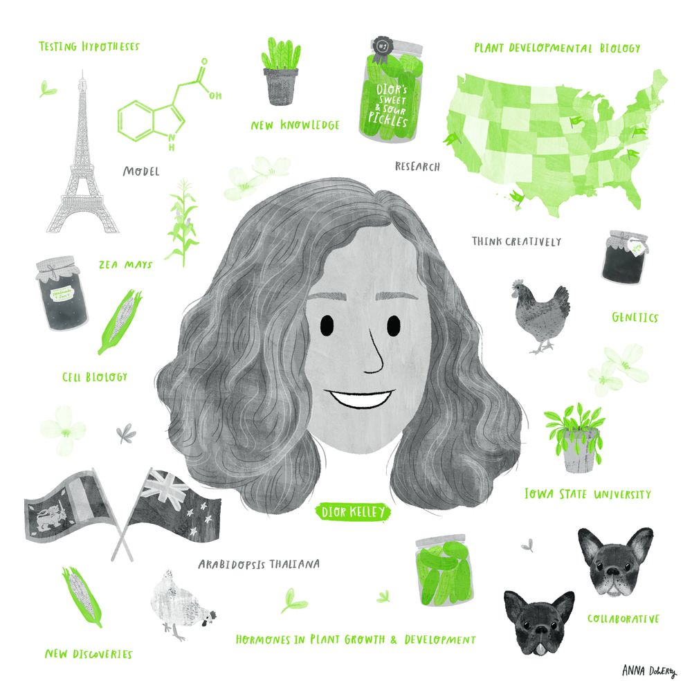
Assistant Professor at Iowa State University. Plant developmental geneticist, loves auxin and roots
Integrative Postharvest Biologist @ucdavisplants. | 🍅🥔 Metabolism & Physiology|Compassionate Excellence| ♥️ Barbados 🇧🇧|♥️ Sea turtle conservation 🐢|
PhD student of Plant-Microbe interactions at LMU Munich, focused on symbiosis with nitrogen-fixing bacteria and receptor-like kinases🌿🍄
Bioinformatician, PhD Candidate | Evolutionary genomics of structural variation and local adaptation in 🌱 Hancock Lab | @mpipz.bsky.social | https://github.com/yykaya
Biochemist based in Aotearoa.
Chemical Biology - Plant hormone signaling - Drug Discovery - Targeted Protein Degradation
Eco-evolutionary biologist 🦎🪰🌸
Interested in the response of organisms to environmental change 🌳🌡️❄️
Postdoctoral researcher at Lund University 🎓
🌾Plant biologist, plant reproduction, seeds, pollen, haploid induction, maize🌽. PI at lab @rdplab.bsky.social in ENS de lyon @ensdelyon.bsky.social / research director at @inrae-france.bsky.social
IBMP is the largest @cnrs.fr center devoted to #plantscience in France | www.ibmp.cnrs.fr
We (@joannakacprzyk.bsky.social and @ucdflowerpower.bsky.social) post one plant memory every day from our survey of plant biologists.
Read the paper here: https://doi.org/10.1002/ppp3.70156
Interested in plant development, cell polarity, Ca+2 and ROS homeostasis, plant cell wall integrity, signalling, protein translation. @Fundacionleloir CONICET Argentina. Currently Editor at @NewPhytologist, @CommsBiology @PlantComms
Prof. @UC Davis ∙structural biologist⚛︎ ubiquitin system aficionado ✾ signaling & sensing mechanisms in plants🌱in vivo interacomes 🎸dad of 2
https://shabek-lab.ucdavis.edu/
🔬 Strongly caffeinated postdoc, ZMBP Tübingen | ✍️ Assistant Features Editor, The Plant Cell @theplantcell.bsky.social
📍 University of Tübingen (DE)
🌐 https://www.pablidopsis.com
Science and innovation for a food and nutrition secure world.
I do plant biology and a few other things at University College Dublin. My team researches plant programmed cell death💀stress responses and mechanisms for plant awareness. Be kind, work hard, have fun🍀🌿🍃🌱
Research group studying the physiology of photosynthesis and its interactions with environmental drivers to improve crop development | Dept. of Plant Sciences, Cambridge
Artur, M.A.S. - Assistant Professor, Seed Resilience Group, WUR.
https://www.mariana-artur.com/ #desiccationtolerance #seedresearch #functionalgenomics #evolution