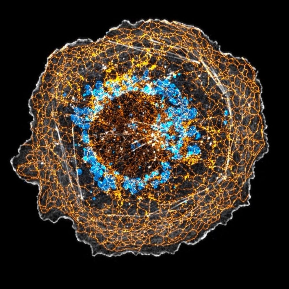
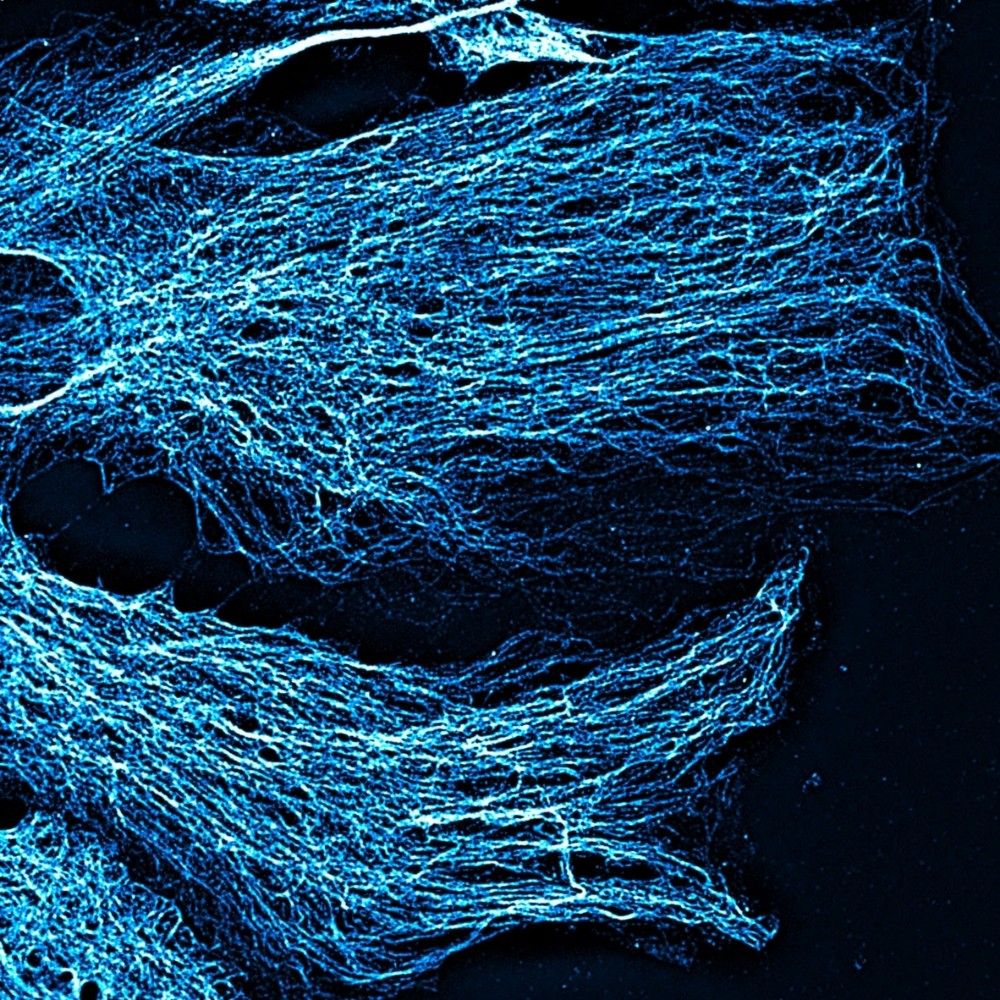
💥 Quick thread on some stunning actin filament swirls🌪️ spotted under platinum replica EM, often forming nest-like structures around caveolae🏐
#ActinAesthetics #ElectronMicroscopy

@tyskalabactual.bsky.social
Interested in how the cytoskeleton controls cell morphology and function; yes to microscopy. https://lab.vanderbilt.edu/tyska-lab/
💥 Quick thread on some stunning actin filament swirls🌪️ spotted under platinum replica EM, often forming nest-like structures around caveolae🏐
#ActinAesthetics #ElectronMicroscopy
Happy Friday! Here to bring a bit of brightness to your day are a few images by Julissa Burgos, one of our graduate students in the Tyska lab here at Vanderbilt. Gorgeous!! 🔬
@yuyi106.bsky.social
@tyskalabactual.bsky.social
#fluorescencefriday #cellbiology
Hey Sam, this one is mStayGold with the following sequence….
www.ncbi.nlm.nih.gov/nuccore/LC75...

This is couple of years old since I rarely have time to sit at a scope these days but I was inspired by the end of year #FluorescenceFriday posts on hear and thought I should drop on y’all my Eye of Sauron, aka a mouse oocyte containing nuclear actin filaments #actin #meiosis #oocytes
20.12.2024 22:56 — 👍 66 🔁 5 💬 0 📌 0
Happy #FluorescenceFriday! These are polarized epithelial cells with microvilli on their apical surface! Captured using the #Nikon NSPARC, and color-coded by depth #actin #microscopy @tyskalabactual.bsky.social
20.12.2024 20:58 — 👍 175 🔁 28 💬 3 📌 1🤩🤩🤩
19.12.2024 23:24 — 👍 1 🔁 0 💬 0 📌 0
This one is for the holiday season
18.12.2024 13:55 — 👍 22 🔁 1 💬 0 📌 0🙏
18.12.2024 02:42 — 👍 0 🔁 0 💬 0 📌 0yes and it’s such a tiny little thing!
17.12.2024 04:04 — 👍 1 🔁 0 💬 0 📌 0
Thought Spef1 one was some ciliary protein? Check out Dr. Rocio‘s (@rtpastrana.bsky.social) poster on how Spef1 shapes epithelial monolayers, no cilia needed 🤩. Tuesday at #cellbio2024, P2439, B46. @vucellimaging.bsky.social
17.12.2024 03:47 — 👍 37 🔁 7 💬 1 📌 1
Have you always wondered how microvilli form? Where DOES that actin come from to form the core bundle? Come to the #BuildingTheCell (Part 1) subgroup tomorrow morning at 8:45 to find out! Room 29C. #CellBio2025
@tyskalabactual.bsky.social
Also, also on Monday at #cellbio2024 I get to present Olivia’s (@operkins.bsky.social) unexpected discoveries on the link between clathrin-mediated endocytosis and the growth of epithelial microvilli 🤯
P1831. B137.
@vucellimaging.bsky.social

Also Monday at #cellbio2024! Julissa (@yuyi106.bsky.social) will present her studies on the role of an I-BAR protein (IRTKS) in the pathogenic mechanism of Enterohemorrhagic E. coli 🦠🧫🔬 P2352. B677. @vucellimaging.bsky.social
16.12.2024 00:08 — 👍 20 🔁 6 💬 2 📌 1
Posters on tuft cells are about as rare as tuft cells, but at #cellbio2024 on Monday, @jen-silverman.bsky.social presents “P1718. Organization of a cytoskeletal superstructure in the apical domain of intestinal tuft cells. B23” Yes! those are gigantic actin bundles 🤯
@vucellimaging.bsky.social
Synchronized filopodial formation for
#FluorescenceFriday 💥
#CellBiology #Microscopy @vucellimaging.bsky.social
Intestinal #organoid goes rogue and flips polarity 🤯
@vucellimaging.bsky.social
#epithelial #cellbiology #microscopy
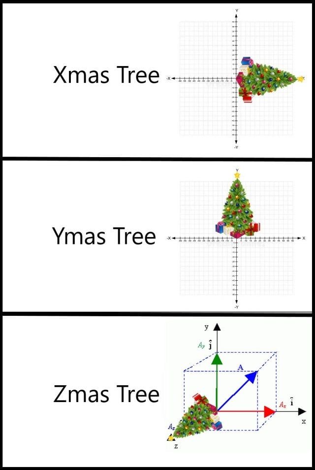
It's that time of year. Confocal + Christmas = Xmas Tree, Ymas Tree, and our favorite Zmas tree. #microscopy #confocal #imaging
11.12.2024 19:04 — 👍 114 🔁 42 💬 1 📌 6As promised, here a fast acquisition of a migrating cell videoed under a microscopy, look those membrane dynamics @ the leading edge !
Bonus: nuclear deformation once the cell rear snaps.
(Fluo nucleus, for the #fluorescencefriday)
@focalplane.bsky.social @cellcommlab.bsky.social #CellMigration 🧪🔬
Highlighting our recent paper on how we think doublecortin (green) contributes to growth cone biomechanics in developing neurons for #FluorescenceFriday
www.cell.com/current-biol...

When you’re imaging and your sample takes a Pikachu!
Having fun playing around with our new #Nikon NSPARC for #FluorescenceFriday
#microscopy #myosin #epithelial #cellbiology #meme
@tyskalabactual.bsky.social
O is for #organoid
@vucellimaging.bsky.social
#microscopy #epithelial #cellbiology
lol
27.11.2024 01:45 — 👍 2 🔁 0 💬 0 📌 0The real IFT…
intra💥filopodial💥transport.
@vucellimaging.bsky.social
#cytoskeleton #biology #microscopy
Starting the week strong! Cheering myself up for the next challenges with 15 min of axonal growth cone dynamic madness. F-actin (cyan) and membrane (magenta) show their dance. One image taken every 10 s.🧠🔬
#MicroscopyMonday #LiveImaging #Neuroscience #Microscopy
Good sampling on this confocal volume of small intestinal tissue showing the apical shag carpet (blue is F-actin, magenta is ZO1)🔬🤩
@vucellimaging.bsky.social
#microscopy #cytoskeleton #epithelial #cellbiology #biology
Looks great, that room has a history of generating awesome data!
23.11.2024 17:59 — 👍 1 🔁 0 💬 1 📌 0
Super proud of pre-Dr. Julissa for going from ND to fundable score with a single revision of her F31 NRSA app🤩🙌🍾🥂🎉🦠🔬
@yuyi106.bsky.social
Extremely rare microtubule staining happening in the lab right now 😭
22.11.2024 21:27 — 👍 8 🔁 0 💬 0 📌 0
First #FluorescenceFriday here on 🦋 Hope you enjoy this cross-section of the intestinal epithelium, highlighting the actin within the #brushborder @tyskalabactual.bsky.social
22.11.2024 21:09 — 👍 34 🔁 6 💬 0 📌 0