Thanks Jonathan, that means a lot to me.
03.04.2025 22:26 — 👍 0 🔁 0 💬 0 📌 0
🙏 (Finally, the perfect use of that emoji)
03.04.2025 22:24 — 👍 1 🔁 0 💬 0 📌 0
Indeed. Biology is merciless to our strengths and weaknesses.
03.04.2025 21:29 — 👍 1 🔁 0 💬 0 📌 0
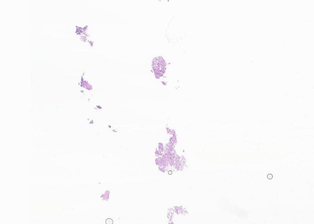
Dx: PD chordoma w/ SMARCB1 loss. I felt so bad I offered to see the patient to apologize but my ortho onc wouldn’t let me lol. Did CK on bx and sure enough, little histiocytoid fragments w/ TINs +. I was humbled, big time. It still haunts me yrs later as I put chordoma in the ddx when I shouldn’t
20.03.2025 05:49 — 👍 2 🔁 0 💬 4 📌 0
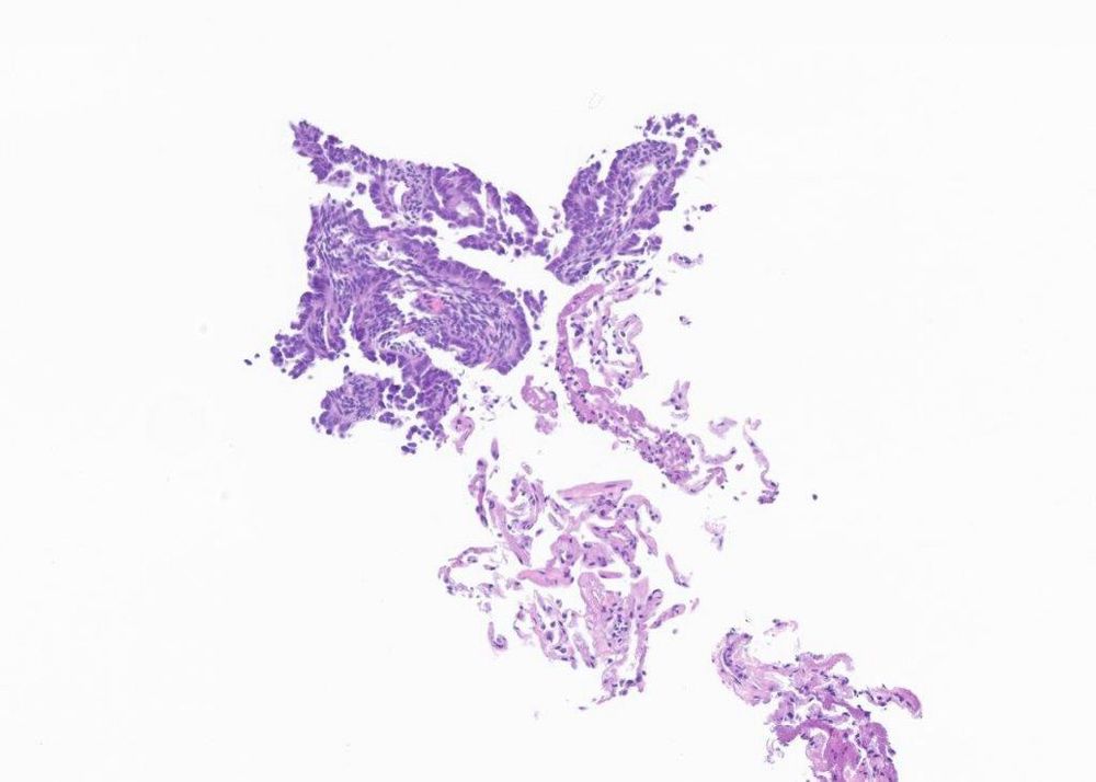
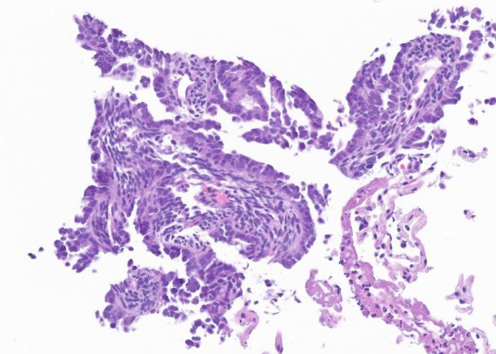
Knee replacement done. Low power. had melting wax look peripherally, w/sheets and large islands of histiocytoid cells with tt necrosis & TINs. CK, EMA +, S100 -, INI1 loss. I called ES. Sent to you guys. Judith emails me it’s brachyury + and my head is spinning.
20.03.2025 05:49 — 👍 1 🔁 0 💬 1 📌 0
Thanks for bringing up. One of my cases was in that paper. I failed twice. Pt was 60s F. Initially, was clinically portrayed as knee TGCT w/ bone erosion. Bx showed synovitis w/ heme, mixed inflam, foam cells and small bits of what looked like histiocytes a/w neuts. I called TGCT.
20.03.2025 05:49 — 👍 1 🔁 0 💬 1 📌 0
I was excited to add ‘modulated’ to my descriptors repertoire 😅
08.03.2025 02:47 — 👍 1 🔁 0 💬 0 📌 0
But what if you miss a cool looking neutrophil on slide 133? Can’t let that happen 😂
07.03.2025 23:01 — 👍 0 🔁 0 💬 0 📌 0
What a saga, I love it!
07.03.2025 22:32 — 👍 0 🔁 0 💬 0 📌 0
Very good to know, thank you.
07.03.2025 21:34 — 👍 0 🔁 0 💬 0 📌 0
Tricky one!
07.03.2025 20:11 — 👍 0 🔁 0 💬 0 📌 0
Okay then, let’s look closer. That material is odd. Spherical ones remind me of tyrosine crystals a bit. The intervening cells are giant cells closely apposed to material, and non descript fibro/myofibro type cells. I’m thinking about pseudogout. Not sure if crystal morph matches though.
07.03.2025 07:37 — 👍 1 🔁 0 💬 1 📌 0
I’m with you, my mistakes are vivid. Some will never be forgotten.
07.03.2025 07:28 — 👍 0 🔁 0 💬 0 📌 0
Calcifying aponeurotic fibroma, schwannoma, pmt? Any stains?
07.03.2025 02:37 — 👍 0 🔁 0 💬 1 📌 0
Excellent case! Never seen one with that pattern
07.03.2025 02:32 — 👍 0 🔁 0 💬 1 📌 0
3rd image made me want STAT6. Then I got instant gratification! Great case, thank you.
14.01.2025 20:30 — 👍 3 🔁 0 💬 1 📌 0
Kal-sp, favor KA. P53, p16, ki67?
14.01.2025 20:27 — 👍 0 🔁 0 💬 2 📌 0
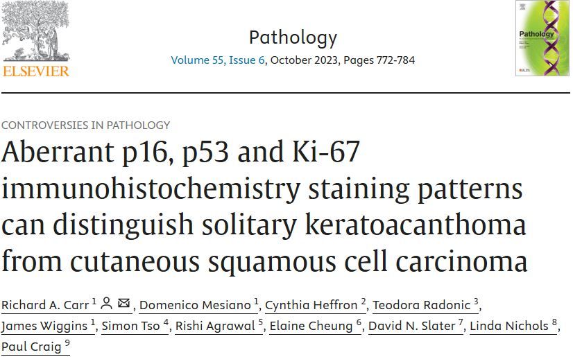
www.sciencedirect.com/science/arti...
Link to the paper that should help everyone diagnose keratoacanthoma with greater confidence.
14.01.2025 08:35 — 👍 18 🔁 4 💬 1 📌 1
Anatomic and Clinical Pathologist, fellowship in #GUPath
Ann Arbor MI
Trustee of American Board of Pathology
College of American Pathologists: The leading organization of board-certified pathologists. #pathologists #Pathsky 🔬
DP fellow@MDACC; Past AP/CP resident@UTSW and SP fellow@BWH
I like my wife and family, bikes, music, dogs, baking bread and soft tissue tumors. Only the last here though.
Pathologist | Postdoc in Image Analysis at Broad Institute
Previously at Erasmus MC, Dana-Farber Cancer Institute
Research areas: GynPath, Premalignant Lesions, Spatial Biology
#PathSky
#Pathology and music. Baldwin-Wallace class of 1978, Yale School of Music 1982 and Columbia College of Physicians and Sugeons 1993. #hematopathology #hemepath
Gastrointestinal pathologist at Emory University
Hematopathologist and Clinical Ethicist at University of Michigan. #medsky #pathsky. Owned by therapy #pug Lyle.
Doctor, pathologist, and medical journalist. This is a personal account.
http://www.mazer.us
https://www.theatlantic.com/author/benjamin-mazer/
UCSD PGY4 Pathology Resident 🔬
Hematopathologist and cytopathologist at Brigham and Women's Hospital
Cardiovascular Pathology, Forensic and Hospital Autopsies.
Pathology Resident - Toronto, Canada
MD PATHOLOGY | PGY 3+4! SPECIAL INTEREST IN BONE AND SOFT TISSUE PATHOLOGY, NEUROPATHOLOGY.
General Pathologist, MD 🔬#CHDVendee La Roche sur Yon 🇫🇷#Francophonepath; interested in #pathology #GIpath #GUpath #Gynpath #Dermpath #Endopath #ENTpath...
Chief Medical Officer and Pathologist, Deciphex/Diagnexia, Pty Ltd
Pathology is my jam 🔬
Here for my two greatest passions in life: science & silly animals
Breast & GYN Pathologist and Cytopathologist
Interim Chair and Director of Anatomic Pathology
Path Residency Program Director
@WVUPathology @WVUMedSchool
Neuropathology trainee @JHU studying brain metastasis with @ashi-w.bsky.social
Assistant Professor at University of Colorado (@CU_pathology) #Hemepath. @StanfordPath
Hemepath Fellowship, AP/CP Residency