"Rising from Chaos" #MicroscopyMonday: an active system of microtubules, kinesin motors and actin filaments.
🔬: Resonant Scanning Nikon AX-R Confocal
🥼: Quang Tran of the Fraden Lab, MRSEC, @brandeisuniversity.bsky.social
🏛️: www.brandeis.edu/science/reso...
📩: info@nesmicroscopy.org
28.04.2025 14:25 — 👍 43 🔁 10 💬 0 📌 3
I love a good olfactory bulb image.
15.04.2025 22:49 — 👍 1 🔁 0 💬 0 📌 0
#microscopy
11.04.2025 15:22 — 👍 2 🔁 0 💬 0 📌 0
🧪
11.04.2025 15:21 — 👍 0 🔁 0 💬 0 📌 0
Choroid plexus is often lost during tissue sectioning and overall anatomy can be difficult if not impossible to reconstruct. Here we observe the vascular anatomy of human choroid plexus as if through a jeweler’s loupe. Sample courtesy of Matthew Schrag and Neil Dani. #FluorescenceFriday
11.04.2025 15:20 — 👍 43 🔁 7 💬 7 📌 2
Thank you to the team at @syGlass for highlighting some of our data. If you haven’t tried working with your data in VR space, you really need to see what you are missing. Syglass is a powerful tool for visualizing and analysis. @vubasicsciences.bsky.social #microscopy #lightsheet #science 🧪
20.03.2025 12:43 — 👍 12 🔁 3 💬 0 📌 0
I'm pleased to report that the board has now heard from NIH that the appointments of these three investigators and similarly situated folks are being extended. Thank you NIH! Delighted that the stellar work there will be continuing!
06.03.2025 22:25 — 👍 397 🔁 92 💬 18 📌 12
🧪 #microscopy #hippocampus #lightsheet
05.02.2025 18:59 — 👍 2 🔁 0 💬 0 📌 0
Here is a closeup on our survey of the #hippocampus from Joe Luchsinger’s mouse sample, using the 15x objective from LifeCanvas Technologies Inc. With #lightsheet imaging you can peel away layers and observe cellular anatomy with stunning detail. How can VNL help you image your sample?
05.02.2025 18:58 — 👍 20 🔁 2 💬 1 📌 0
For #FluorescenceFriday, a side-by-side comparison of 5xFAD hemibrains at 6 months showing how Aβ plaques can be modulated not just by sex but also by the transgene parentage.
More details from our recent paper where we reported the transgene inheritance effect in 5xFAD mice 👇
31.01.2025 15:27 — 👍 7 🔁 3 💬 0 📌 0
It’s always great to get lucky with a sample😆
01.02.2025 20:00 — 👍 1 🔁 0 💬 0 📌 0
Thank you 🙏
28.01.2025 23:41 — 👍 1 🔁 0 💬 0 📌 0
Just a gorgeous #Purkinje neuron all by itself in the cerebellum. Only with #lightsheet #microscopy on a whole #cleared sample could you ever hope to catch a lone reporter expressing cell in it’s entirety. #science 🧪
28.01.2025 23:39 — 👍 210 🔁 43 💬 9 📌 6
#microscopy
20.01.2025 18:18 — 👍 0 🔁 0 💬 0 📌 0
#microscopy #lightsheet
20.01.2025 03:38 — 👍 1 🔁 0 💬 0 📌 0
Definitely need to try this. Has anyone here used this on lightsheet meso scale data? @microscopy 🧪
20.01.2025 03:04 — 👍 13 🔁 2 💬 0 📌 0
Hi Julia! My lab uses the smartSPIM from Life Canvas Technologies (Boston, Massachusetts) for imaging. This was cleared using their SMARTBATCH+
18.01.2025 16:32 — 👍 1 🔁 0 💬 0 📌 0
Thank you!🙏
18.01.2025 16:06 — 👍 1 🔁 0 💬 0 📌 0
Ugh. Bsky is applying aggressive video compression and lots of detail is being lost.
18.01.2025 00:40 — 👍 0 🔁 0 💬 0 📌 0
🧪
17.01.2025 23:15 — 👍 1 🔁 0 💬 0 📌 0
Ok here’s a first go at looking at the previously acquired sample in 3D space. Still some work to do before we have a final product. 🧪
17.01.2025 20:44 — 👍 31 🔁 2 💬 6 📌 0
You know what it takes to get images like these! 😅
16.01.2025 20:42 — 👍 1 🔁 0 💬 0 📌 0
Thank you! I’m still rebuilding the entire volume.
16.01.2025 20:41 — 👍 2 🔁 0 💬 1 📌 0
Thank you Sunny! Transgenic expression is always the most photogenic!
16.01.2025 19:58 — 👍 1 🔁 0 💬 1 📌 0

Getting closer… 🧪
16.01.2025 19:57 — 👍 26 🔁 5 💬 2 📌 0
It’s a flourescent protein
16.01.2025 18:38 — 👍 0 🔁 0 💬 0 📌 0
These are tdTomato expressing neurons in a cleared, intact mouse brain.
16.01.2025 18:22 — 👍 0 🔁 0 💬 1 📌 0
🧪
16.01.2025 17:04 — 👍 2 🔁 0 💬 1 📌 0
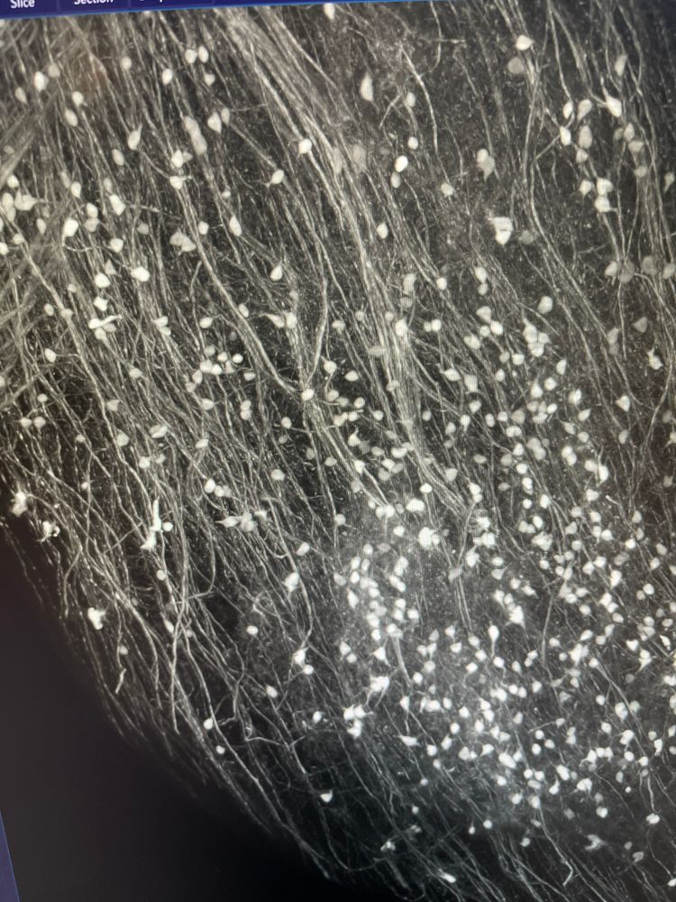
Image preprocessing. This is from the acquisition in my previous post. Final image will be gorgeous!
16.01.2025 17:03 — 👍 20 🔁 0 💬 3 📌 0
Green fluorescent protein is being illuminated by a sheet of laser light. The protein is found inside of a population of neurons which together constitute a putative brain circuit. The sample is an intact mouse brain which has been chemically modified to be optically transparent.
13.01.2025 18:46 — 👍 1 🔁 0 💬 0 📌 0
The Department of Biotechnology and Biosciences of the University of Milano-Bicocca
Hi 👋 I'm a postdoc in the #Neuroimmunology and #Imaging group at the @dzne.science Bonn 🧪🔬 Passionate about #ComputationalNeuroscience 🧠💻 and #NeuralModeling 🧮
🌍 fabriziomusacchio.com
👨💻 github.com/FabrizioMusacchio
🐘 sigmoid.social/@pixeltracker
Psychologist | Assistant Professor (PhD) in Criminology at UCLM (Spain)| Criminology Research Center 🎓🔍 | Research Group on Victimology and Psychopathology of Childhood and Adolescence | Passionate about human behavior, plot twists & dark minds 🎬🕵️♀️
I'm a scientist studying resilience w/ children & families who experience adversity, like homelessness & loss.
RG: www.researchgate.net/profile/J-J-Cutuli/publications
Substack: https://joewillard.substack.com/
Neuroscientist at the Paris Brain Institute / www.dejuansanzlab.org / ERC / FENS-Kavli Scholar / Young Academy of Spain / CNRS
PhD - Senior Research Assistant in Stapornwongkul Lab (IMBA, Vienna).
Cell and developmental biologist.
Passionate bioimager and photographer.
Institute of Molecular Biology, BAS
#DNArepair #Replication #Genomeintegrity #Cancer #Anticancerdrugs #Microscopy
Leibniz Institute: Hans Knöll Institute
Jena
The research group Applied Systems Biology is concerned with the mathematical modeling and computer simulation of infection processes caused by human-pathogenic fungi.
Kirschstein-NRSA Postdoctoral Research Fellow at UVA studying B1 cells in various diseases w focus on fibrosis. Likes making tools. Flow cytometry + microscopy + data wrangler.
Wannabe polymath.
Leftist/anti-authoritarian hot takes are my own.
Neurobiologist, osmo-electro-diffusion & other stuff. Views are my own.
Academic Cardiac and Mobile Anaesthetist | University of Melbourne and Medical School | Medical Director of Sleep Dentistry Services | Day Care Anaesthesia Special Interest Committee
🌐 www.sleepdentistryservices.com.au
📞 0429 058 878
The official account for the Life Science Solutions division of the Nikon Healthcare Business, with a focus on biological microscopy products and services for the life science, biotech/pharma, and clinical laboratory markets.
This is the official account for the Society for Developmental Biology. Skeets on all the latest Dev Bio news, meetings and science! www.sdbonline.org
Neurotox postdoc | Tjalkens laboratory @CSU
adamschuller.com
Helping people reconnect with Nature - custodians, not always customers | Some humour & comments, too | My website https://tinyurl.com/Henricusp | I am lead editor of NAEE Journal www.naee.org.uk & Wildlife Australia magazine
Prof at Saarland. NLP and machine learning.
Theory and interpretability of LLMs.
https://www.mhahn.info
Canada Research Chair. Misfolding proteins, chaperones and cholinergic function in neurodegeneration. Interested in reproducibility, translational research and open science. ORCID: 0000-0002-3028-5778
I also post pics of sunsets and bikes.