I have some BIG NEWS to share today!
Today I begin my next adventure as
Chief Editor of Nature Biomedical Engineering!!
(a thread) @natmethods.nature.com @natbiomedeng.nature.com @natureportfolio.nature.com

@fspasqualini.bsky.social
I have some BIG NEWS to share today!
Today I begin my next adventure as
Chief Editor of Nature Biomedical Engineering!!
(a thread) @natmethods.nature.com @natbiomedeng.nature.com @natureportfolio.nature.com
We’re hiring! 🚀 Junior Lab Tech / PhD student (cell & mol-bio) to push our Calipers tech into 3D #gastruloids. Must already have Italy work permit (sorry, can’t sponsor). Love stem cells + CRISPR? DM or send CV by 31 Aug 2025. #hiring #StemCells
13.07.2025 08:01 — 👍 2 🔁 1 💬 0 📌 0#CALIPERSv2 🚀 preprint is locked—journal submission next. Final polish, more hiPSC reference lines, and fresh light-sheet data. Still, a great collaboration led by @moisesdisante with the team @LabPhysiology and the amazing @berterolab.bsky.social and @florianjug.bsky.social group members! 1/N
08.07.2025 09:36 — 👍 8 🔁 4 💬 1 📌 2A tour de force that elevates the #CALIPERS technology to a whole new level! A must read for all iPSCs, cell cycle, and developmental biology aficionados.
08.07.2025 10:05 — 👍 4 🔁 2 💬 0 📌 1Everything is open-science here, links in the pre-print, but reach out if you don't want to wait for the final repositories to be updated. www.biorxiv.org/content/10.1...
08.07.2025 09:36 — 👍 2 🔁 0 💬 0 📌 0By tracking rare cycling hiPSC-derived cardiomyocytes, we can monitor cell proliferation (useful in regeneration) and multinucleation and endoreplication (useful for assessing maturation) as well as structure and function integration #CCAwarePhenotyping #CCAwareRegenerativeStudies #MethodInACellLine
08.07.2025 09:36 — 👍 3 🔁 1 💬 1 📌 0New light-sheet capture of 4-color hiPSC-cardioids reveals compaction before calcium transients initiation. 90 % exit cycle by d4.5 → maturation on cue. #CCAwarePhenotyping #MethodInACellLine
08.07.2025 09:36 — 👍 2 🔁 0 💬 1 📌 0Safe-harbor genome editing ensures expression across differentiated lineages, and WTC-11 genetic background means full compatibility with the
@alleninstitute.org Cell Collection: the largest repository of GFP/RFP-based hiPSC reporter lines www.allencell.org/cell-catalog...
WTC11-based hiPSCs reporter lines. We provide a 4-color CALIPERS hiPSC line with sensors for actin, calcium, and the cell cycle, as well as a blank canvas line to add your favorite sensors. CALIPERS is a #MethodInACellLine!
08.07.2025 09:36 — 👍 2 🔁 0 💬 1 📌 0Phase-locked motility
Each nucleus gets a %-cell-cycle score that rides with its mask and trajectory. Compare shape or speed by cycle progress, not frame time. Watch HaCaTs sprint through physical space and cell cycle (CC) phases! #PhaseLockedMotility #CCAwarePhenotyping
Proposed solution: To overcome this, we integrated a spectrally re-engineered FUCCI variant, open-source analysis software, and four-color human stem cell reporter lines into CALIPERS: a method for Cell-cycle-Aware Live-cell Imaging in Phenotyping and Regeneration Studies.
08.07.2025 09:36 — 👍 5 🔁 1 💬 1 📌 0Problem: Cell cycle progression, migration, and proliferation shape development and regeneration, but simultaneous live-cell imaging remains challenging as conventional fluorescent cell cycle indicators (FUCCI) monopolize the green and red channels used by most structural and functional biosensors.
08.07.2025 09:36 — 👍 2 🔁 0 💬 1 📌 0#CALIPERSv2 🚀 preprint is locked—journal submission next. Final polish, more hiPSC reference lines, and fresh light-sheet data. Still, a great collaboration led by @moisesdisante with the team @LabPhysiology and the amazing @berterolab.bsky.social and @florianjug.bsky.social group members! 1/N
08.07.2025 09:36 — 👍 8 🔁 4 💬 1 📌 2The preprint is now available! Check it out... and... if you want to repost, why not 👇the post down here👇, so others get the bioRxiv link right away? Oh, and... 𝐭𝐡𝐚𝐧𝐤𝐬! 🙏
doi.org/10.1101/2025...
bsky.app/profile/flor...
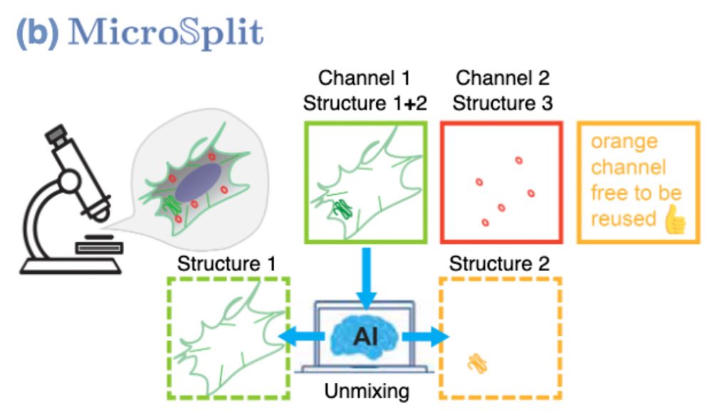
Schematic overview on how MicroSpit operates. It can split superimposed structures in fluorescent image channels by using a suitably trained AI.
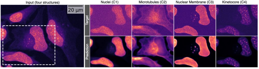
Concrete example for how MicroSplit unmixes four superimposed structures.
Immagine you could image two cellular structures in the same fluorescent channel and still reliably get them separated afterwards…
What would you do with this?
Now… what would you do if that also worked with 4 structures at once? 👇 #MicroSplit #preview🧵
🔬🎤 As pre-announced a few days ago... please let us proudly present to you: 𝑴𝒊𝒄𝒓𝒐𝕊𝒑𝒍𝒊𝒕 - your ticket to imaging more, imaging more gentle, and/or imaging more efficient. 🔬
doi.org/10.1101/2025...
Like ❤️, repost 🔂, and most importantly... please send feedback ✉️ our way! 🙏
You are too kind! And we definitely couldn't have done this without the help of your lab and @florianjug.bsky.social's!
10.02.2025 08:46 — 👍 2 🔁 0 💬 0 📌 0And @berterolab.bsky.social joined us here on Bluesky, give him and his amazing lab a Follow!
09.02.2025 10:22 — 👍 2 🔁 0 💬 0 📌 0Hello, Bluesky! ☀️
We fund frontier research in Europe—bold ideas, unexpected discoveries and science that shapes the future. So it’s only fitting we’ve landed here. Sorry for being late.
Follow us for updates on ERC funding, research policy, and our grantees' discoveries.
HYDRA supports fluorescence microscopy, enabling cell-cycle and cytoskeletal analyses with FUCCIplex sensors using CALIPERS (biorxiv.org/content/10.1...). From cell proliferation to mitotic spindle dynamics, this method works with cutting-edge imaging and image-analysis modalities.
17.01.2025 16:14 — 👍 2 🔁 1 💬 1 📌 0And works with organoids and light sheet microscopy as well!
23.01.2025 20:02 — 👍 6 🔁 4 💬 0 📌 0And works with organoids and light sheet microscopy as well!
23.01.2025 20:02 — 👍 6 🔁 4 💬 0 📌 0I'm happy to announce a postdoctoral position is available in my lab. We have lots of great data to analyze with respect to transposons & epigenetics and cool biological questions to answer.
Please reskeet and share.
Applications for our Quantitative Imaging: From Acquisition to Analysis 2025 course (Mar 24-Apr 8) are due on Jan 31! Come immerse yourself in microscopy and image analysis with me, @talleylambert.bsky.social, @bethcimini.bsky.social, @florianjug.bsky.social & Hunter Elliott! Please RT
21.01.2025 19:09 — 👍 45 🔁 27 💬 0 📌 0This is wild. I made a silly little post yesterday. Richard responded with a joke, and then "Cassandra" hopped in the thread. I thought her responses were weird, but then Richard caught on and posted this. Clever way to catch a bot!
20.01.2025 20:30 — 👍 1099 🔁 271 💬 30 📌 31And here is pre-print 2/2! *HYDRA: HYdrogel Dispensing with Robotic Automation* - a scalable solution for drug testing. Great work by 1st author
@eloisa_torchia and team with help from the labs of
@sgabriele.bsky.social Elisa Cimetta and Johan Lind biorxiv.org/content/10.1...
With
@florianjug.bsky.social's team, we developed an open-source FUCCIphase plugin to calculate CC phase percentages, thus enabling CC-aware morphology/motility analyses in live cells.
In conclusion, HYDRA is a cost-effective, HTS-compatible solution that integrates seamlessly into existing pharma workflows, ideally making early preclinical drug testing more predictive. Thoughts, questions, or collaborations? Let’s connect.
17.01.2025 16:14 — 👍 0 🔁 0 💬 0 📌 0HYDRA supports fluorescence microscopy, enabling cell-cycle and cytoskeletal analyses with FUCCIplex sensors using CALIPERS (biorxiv.org/content/10.1...). From cell proliferation to mitotic spindle dynamics, this method works with cutting-edge imaging and image-analysis modalities.
17.01.2025 16:14 — 👍 2 🔁 1 💬 1 📌 0