Going to have to carve some time out for this!
10.02.2026 01:39 — 👍 1 🔁 0 💬 0 📌 0
When your cat is obsessed with #PuppyBowl. As a special needs adoptee herself, she’s a huge advocate.
09.02.2026 01:17 — 👍 6 🔁 0 💬 0 📌 0
Tim Mosca, Thomas Jefferson University
Office of University Interdisciplinary Programs
I am very excited to visit NC State this weekend and give a seminar on Monday! I'll be in Raleigh from Saturday to Tuesday so I'm joyfully accepting all running trail suggestions, dining ideas, cool places to sit and write 6 R01s, and any friends who want to say Hi!
gga.ncsu.edu/event/tim-mo...
07.02.2026 02:35 — 👍 20 🔁 2 💬 4 📌 0

Microscopy image of a kidney only showing the highly branched arterial tree in magenta against a black background.
The beauty of the kidney arterial tree for this #FluorescenceFriday. Evans Blue dye labels the vasculature. Image courtesy of postdoc Sarah McLarnon.
06.02.2026 22:16 — 👍 80 🔁 13 💬 0 📌 0
Hope so too as I assume the problem will only get worse as deadlines approach. Still no progress with mine.
14.01.2026 00:11 — 👍 0 🔁 0 💬 0 📌 0

Announcement of the show and a view of last year's crowd
Great things still happen all around us. Each your UNC Biology's Bob Goldstein and Art's Beth Grabowski co-teach ARTS/BIOL 409, that brings together artists and scientists to explore an intersection of two disciplines. It culminated this year with a remarkable show Friday 1/n
11.01.2026 23:04 — 👍 40 🔁 8 💬 1 📌 2
That’s great news! 🤞
10.01.2026 00:17 — 👍 0 🔁 0 💬 0 📌 0
Ugh, I’m sorry. Not much better here. My request is still somewhere in the ether with an auto reply date of January 20th for resolving issues. 😭
09.01.2026 21:39 — 👍 0 🔁 0 💬 1 📌 0
Pro tip: NEVER log into My NCBI with more than one 3rd party option. It creates a whole new account that can’t be deleted. I apparently did this once with the ORCiD login (as opposed to normal eRA login) and now I can’t link my ORCiD ID to my SciENcv. I have to ask the Help Desk to merge accounts. 🫠
09.01.2026 17:43 — 👍 6 🔁 4 💬 2 📌 1
Introducing Cyclically Multiplexed Expansion Microscopy (Cy-ExM): a workflow for 3D nanoscale, high-plex imaging in whole cells. Cryo-preserved ultrastructure + iterative labeling + expansion microscopy → 20 targets in one dataset with ~70 nm lateral resolution.
www.biorxiv.org/content/10.6...
02.01.2026 18:18 — 👍 30 🔁 8 💬 2 📌 1
Thanks so much, Rejji!
31.12.2025 21:41 — 👍 1 🔁 0 💬 0 📌 0

Image of a developing kidney collecting duct system in yellow with innervating nerve fibers in cyan.

Image of a sectioned ganglion with many colors highlighting the cell bodies and nuclei.

Image of a developing kidney showing nephron progenitors in yellow and collecting duct system in blue.
It’s certainly been a year, but I’m incredibly grateful that earlier in 2025 I was promoted to Associate Professor with tenure! Huge thanks to you all for promoting our work here and on the other site, for talk invites, for writing letters of support, and to the lab for all their contributions. 🥂
31.12.2025 14:19 — 👍 30 🔁 3 💬 1 📌 0
Unfortunately the drop in ESI funding rates is not paired with changes in funding required to get tenure. It’s not just the next three years of our labs at risk, but the rest of our careers. We are also the cohort that started in the COVID pandemic- it’s been nothing but “unprecedented times”…
19.12.2025 02:16 — 👍 23 🔁 10 💬 0 📌 1

Most transcriptomics studies sample ~0.001% of a mouse. Which region do we choose? Often: we guess.
Meet DISCO-seq 🤗 flipping the order: RNA-preserving clearing → whole-organ/whole-body 3D imaging → pick ROIs in 3D → unbiased scRNA-seq.
Preprint: www.biorxiv.org/content/10.6...
18.12.2025 14:44 — 👍 25 🔁 8 💬 1 📌 4

title, authors and abstract from link
In a paper out this month in @jcb.org we present evidence implicating intrinsically disordered regions (IDRs) as key to junctional assembly and function 2/n 🧪
pubmed.ncbi.nlm.nih.gov/41099653/
14.12.2025 15:00 — 👍 23 🔁 7 💬 1 📌 1
Our new work Cilia.io is out on the Rxiv. Ece Atayeter and Jason Ho trained an ML computer vision transformer for detection and quantification of cilia morphology and dynamics. We will provide a GUI soon. Another great collaboration w/ @jbwallingford.bsky.social
13.12.2025 19:14 — 👍 12 🔁 1 💬 0 📌 2

Enhancer-directed gene delivery for digit regeneration based on conserved epidermal factors
Limb loss remains a significant clinical challenge, but regenerative medicine approaches such as gene therapy offer a promising strategy to trigger endogenous regeneration programs. Optimal vector con...
🚨 Preprint Friday! 🚨
Thrilled to highlight this collab with David Brown at Duke @cutitoutdave.bsky.social and Ken Poss at UW Madison/Morgridge @kenposs.bsky.social. By comparing 3(!!) regenerating models we identify a conserved role for Sp-family TFs in appendage regeneration (and more).....🧵
05.12.2025 20:18 — 👍 23 🔁 9 💬 2 📌 0
I think this is it. While this isn’t a change for some ICs in terms of percentile cut offs, how are these additional criteria, that were not always taken into account before, going to be balanced? What is their weight in decisions on funding?
22.11.2025 03:40 — 👍 0 🔁 0 💬 0 📌 0


Durham, NC about 10 minutes ago
13.11.2025 03:08 — 👍 0 🔁 0 💬 0 📌 0
Flashback #FluorescenceFriday to some of the first light-sheet imaging we were doing. Video shows the collecting duct system of the developing kidney labeled with a pan-cytokeratin antibody. Courtesy of former talented technician Deanna Hardesty.
07.11.2025 16:43 — 👍 78 🔁 22 💬 1 📌 1

🆕 KidneyCure is committing up to $6 million over three years to help eligible NIH R01 applicants ranked in the top 15% continue their kidney research with a $100K award.
Learn more and explore every KidneyCure opportunity: https://bit.ly/kcgrantsfunding
16.10.2025 19:00 — 👍 2 🔁 1 💬 0 📌 0
Yeah, this year is chaos for study sections. Mine was scheduled for last week. We got a email from CSR communications the night before telling us it was postponed and not to join the scheduled meeting.
14.10.2025 20:16 — 👍 2 🔁 1 💬 0 📌 0

We present multi-immersion Oblique Plane microscope (miOPM), a light-sheet platform that can be adapted to a wide range of applications, from sensitive live cell imaging to imaging organs and cleared tissues.
www.biorxiv.org/content/10.1...
06.10.2025 17:52 — 👍 123 🔁 44 💬 5 📌 2

Figure from our review paper showing various fluorescence microscopy images of cells and structures of the kidney
Sharing this excellent review now out from postdoc Sarah McLarnon! Lots of useful info on 3D imaging, quantification of imaging data, and how this has been applied to the kidney. And since it's #FluorescenceFriday, sharing a figure with our own imaging data!
www.sciencedirect.com/science/arti...
03.10.2025 23:29 — 👍 38 🔁 8 💬 0 📌 0

RhoA (blue) and the actin cytoskeleton (magenta) are shown in a set of primary microglia.
For #FluorescenceFriday, RhoA (blue) and the actin cytoskeleton (magenta) are shown in a set of primary microglia 🔬 #Neuroscience #Microscopy
03.10.2025 13:07 — 👍 83 🔁 16 💬 4 📌 1
My entry for today’s #FluorescenceFriday: a pupal #Drosophila testis with muscles expressing
🔵 lifeact &
🔴 RFP-nls
Honored & grateful to receive an honorable mention at @healthcare.nikon.com Nikon Small World 🌍🔬✨
🔗 www.nikonsmallworld.com/galleries/20...
#NikonSmallWorld #Microscopy #ScienceArt
03.10.2025 09:06 — 👍 229 🔁 44 💬 12 📌 2

Zoomed up image of the collecting ducts of an adult mouse kidney. The collecting ducts are labeled with an AQP2 antibody and are green. They look like long green squiggles.
And a zoomed view of the intricate collecting system.
26.09.2025 17:51 — 👍 8 🔁 1 💬 0 📌 0

Image of a fluorescently labeled adult mouse kidney showing AQP2 staining of collecting ducts and connecting segment in green and alpha SMA staining of the arterial tree in magenta. The collecting ducts look like squiggly branches.
For this #FluorescenceFriday, a gorgeous image of an adult mouse kidney labeled with AQP2 and alpha SMA antibodies. AQP2 (green) marks the collecting duct and distal connecting segment while SMA marks the arterial tree. Courtesy of talented postdoc Sarah McLarnon.
26.09.2025 17:51 — 👍 116 🔁 32 💬 5 📌 0
Exploring how hyaluronan dynamics shape developmental timing, aging, and disease. 💧
OSF: https://osf.io/sd5g4/
Hi, I am a scientist working on epithelial Morphogenesis @IBDM Marseille. Passionate about Development, Shape, Evolution, and turning pretty pictures into numbers
Beer, Radiation, Music, and Lab Safety...not necessarily in that order. Views are my own.
#bloodbrainbarrier 🩸🧠🚧
#neuroscience 🧫🔬
Antibody developer at SySy Antibodies.
A human who’s kind of bad at social media. Stanford Neuro PhD. Postdoc in the Hill lab✨ @OHSU. Science is for everybody!! Mama🫶
(she/her)
Virginie Hamel & Paul Guichard Lab at University of Geneva
#cryoEM/ET❄️ and #UExM ⚗️ #ExpansionMicroscopy #TeamTomo
Genève, Suisse 🇨🇭
https://mocel.unige.ch/research-groups/guichard-hamel/overv
Postdoc at Duke Univ Cell Bio
Interested in cell communication in a multicellular society, morphogenesis, and epithelial homeostasis.
🇿🇦🇳🇿🇺🇸Neuroscientist/amateur virologist. Professor and Director of the Molecular Biology Graduate Program, University of Utah. Molecular mechanisms of memory, repurposed transposons, and virus-like intercellular signaling. Personal views.
www.shepherdlab.org
Focused on innate immune mechanisms governing pathogenesis of disease and on NK-cell based therapies for disease. Opinions expressed are Dr. Waggoner's alone and not on behalf of lab staff or employer.
Professor of Animal Physiology @uni-muenster. Cavefish enthusiast, EvoDevo aficionado. Exploring starvation resistance & overeating in cavefish (and myself: intermittent fasting devotee and food lover).
Mourning the days when all we talked about was font choice, reference styles and glam humping.
Computational Biology @ETH: data-driven modeling & simulation of emerging phenomena in development & disease https://bsse.ethz.ch/cobi https://youtube.com/@cobi-ethz
Policy Professor, Ford School, University of Michigan.
Irish immigrant. Administrative burdens guy.
Free newsletter, Can We Still Govern?: https://donmoynihan.substack.com
Prof. @ucsantabarbara.bsky.social - Runs a lab slslab.org - Works on computation, neuroscience, behavior, vision, optics, imaging, 2p / multiphoton, optical computing, machine learning / AI - Blogs at labrigger.com - Founded @pacificoptica.bsky.social
Developing integrated technological platforms to study the molecular mechanisms that drive processes in development and disease.
@ Fiolka Lab, UT Southwestern
PhD student in neuroscience @ VUW 🇳🇿
environmental enrichment, neuromorphology, synapses, mass spec proteomics
https://github.com/stephaniejh
Developmental biologist | Professor at Indiana University Department of Biology | eye development, pattern formation, gene regulation
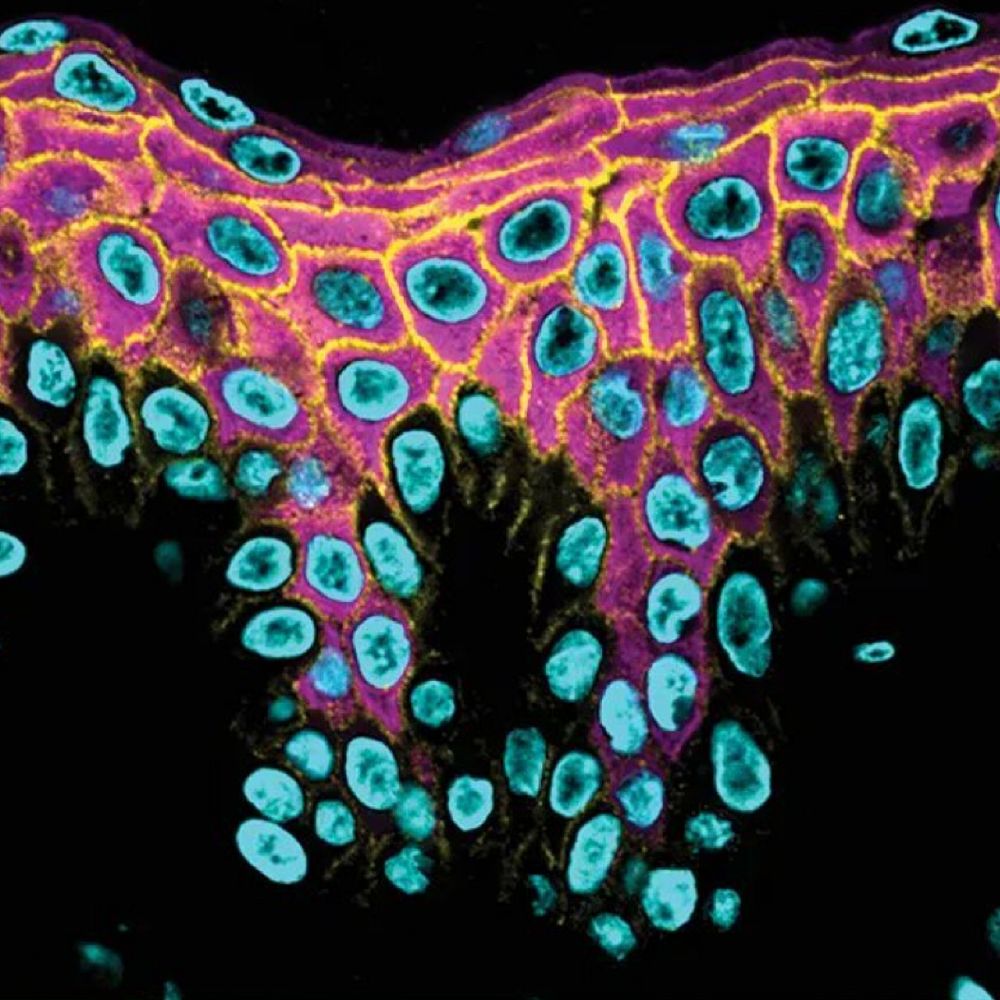
Illuminating keratinocyte cell biology in the Dept of Dermatology at Univ of Washington to understand epidermal differentiation/integrity & their compromise in skin disease. PI: Cory Simpson, MD/PhD, dermatologist specializing in rare blistering disorders.
Physician (IM, EM, Pulm), Scientist. Dancer, Seamstress, Apprentice Balcony Gardener, Booklover, Foodie (more eating than cooking), Painter (badly), Traveller, Gamer. Living with Crohn's Disease.
Swiss (but not a fan of chocolate).