Thanks Henry for your hard work!! It was fun indeed :)
13.09.2025 09:28 — 👍 0 🔁 0 💬 0 📌 0
Enjoy guys!! 🍾🥳
11.09.2025 21:21 — 👍 5 🔁 0 💬 0 📌 0
We currently have open positions for PhD and Postdocs! Interested in learning fUS: please apply!
brainwidenetworks.uni-goettingen.de/open-positio...
11.09.2025 20:12 — 👍 5 🔁 3 💬 0 📌 0
Big thanks to our institutions and funding sources for the support—and to everyone on the team for making this discovery possible! 🙏✨ @mbexc.bsky.social @mpiforbi.bsky.social @mcgill.ca @dfg.de
11.09.2025 20:12 — 👍 1 🔁 0 💬 1 📌 0

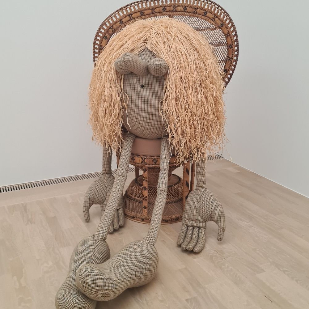
In summary, visual objects refine population-level head-direction coding in postsubiculum, potentially helping the brain’s internal compass anchor to external cues. Whether this extends to other types of spatially tuned neurons remains an exciting open question!
8/
Illustration: Dorothea Laurence
11.09.2025 20:12 — 👍 5 🔁 2 💬 2 📌 0

To test if this effect was specific to objects, we presented two landmarks to the mouse: an object picture or a scrambled version. The boost occurred only with the object!
7/
11.09.2025 20:12 — 👍 1 🔁 0 💬 2 📌 0

At the population level, head-direction cells form a ring attractor. Cells aligned with an object’s direction were boosted, while others were inhibited—showing that objects refine the brain’s internal compass.⚡🧭 A model confirmed the effect when adding an untuned input to the attractor network.
6/
11.09.2025 20:12 — 👍 3 🔁 1 💬 1 📌 0

We then asked: How are visual signals integrated with spatial ones? We teamed up with @apeyrache.bsky.social. Mice were recorded in PoSub while exploring an arena with a landmark, then head-fixed for visual stimulation. Both head-direction cells and fast-spiking interneurons preferred objects!
5/
11.09.2025 20:12 — 👍 2 🔁 0 💬 1 📌 0
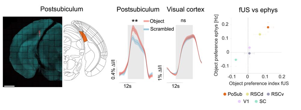
To our surprise, spatial navigation areas—not visual cortex—responded strongest to objects! We replicated this in awake and anesthetized mice and confirmed it with electrophysiology. Postsubiculum (PoSub), a hub of the head-direction system, was the top hit! 🎯
4/
11.09.2025 20:12 — 👍 11 🔁 2 💬 1 📌 0

This project began with a paradox: Mice can see objects, yet no dedicated object areas like those in primates had been found. Inspired by early human fMRI studies, we used an unbiased functional ultrasound (fUS) screen to look beyond the visual cortex.
3/
11.09.2025 20:12 — 👍 2 🔁 0 💬 1 📌 0
This was a true team effort, led by the brilliant Domique Siegenthaler, in collaboration with Stuart Trenholm and @apeyrache.bsky.social ! 🙌
2/
11.09.2025 20:12 — 👍 1 🔁 0 💬 1 📌 0

Thrilled to share that our work is now published in Science! ✨
We found a preference for visual objects in the mouse spatial navigation system where they dynamically refine head-direction coding. In short, objects boost our inner compass! 🧭
www.science.org/doi/10.1126/...
🧵1/
11.09.2025 20:12 — 👍 175 🔁 72 💬 8 📌 6

To test if this effect was specific to objects, we presented two landmarks to the mouse: an object picture or a scrambled version. The boost occurred only with the object!
7/
11.09.2025 19:28 — 👍 0 🔁 0 💬 0 📌 0

At the population level, head-direction cells form a ring attractor. Cells aligned with an object’s direction were boosted, while others were inhibited—showing that objects refine the brain’s internal compass.⚡🧭 A model confirmed the effect when adding an untuned input to the attractor network.
6/
11.09.2025 19:28 — 👍 0 🔁 0 💬 1 📌 0

We then asked: How are visual signals integrated with spatial ones? We teamed up with @apeyrache.bsky.social. Mice were recorded in PoSub while exploring an arena with a landmark, then head-fixed for visual stimulation. Both head-direction cells and fast-spiking interneurons preferred objects!
5/
11.09.2025 19:28 — 👍 0 🔁 0 💬 1 📌 0

To our surprise, spatial navigation areas—not visual cortex—responded strongest to objects! We replicated this in awake and anesthetized mice and confirmed it with electrophysiology. Postsubiculum (PoSub), a hub of the head-direction system, was the top hit! 🎯
4/
11.09.2025 19:28 — 👍 0 🔁 0 💬 1 📌 0

This project began with a paradox: Mice can see objects, yet no dedicated object areas like those in primates had been found. Inspired by early human fMRI studies, we used an unbiased functional ultrasound (fUS) screen to look beyond the visual cortex.
3/
11.09.2025 19:28 — 👍 0 🔁 0 💬 1 📌 0
This was a true team effort, led by the brilliant Domique Siegenthaler, in collaboration with Stuart Trenholm and @apeyrache.bsky.social ! 🙌
2/
11.09.2025 19:28 — 👍 0 🔁 0 💬 1 📌 0
Neuroscientist interested in sensory processing and brain-wide circuit dynamics.
Natural behavior, brain evolution, & ecological change, with a focus on wild 🐭 and 🦎. Currently a Postdoc @mpibrain.bsky.social; PhD from @harvardmcb.bsky.social. he/him
felixbaier.github.io
Electrophysiologist-ProteinEngineer-RhodopsinEnthusiast-2PVoltageImager
Comp. Cog. Neuro, Imaging & Neurphysiology, Epilepsy | MNI, McGill U.
Systems neuroscientist | Working out how the 🧠 generates 🌐 to find its 🧭 | Lecturer (Asst Prof) at the University of Manchester | Big fan of ancient things 🏺📜
Neuroscientist. Interested in activity patterns in the cortex, unaffordable guitars and mexican food. Worked at Yale University, University of Amsterdam and ESPCI Paris
Neuroscientist at European Neuroscience Institute. Postdoctoral researcher.
I post mainly about Neuroscience, Machine Learning, Complex Systems, or Stats papers.
Working on neural learning /w @auksz.bsky.social CCNB/BCCN/Free University Berlin.
I also play bass in a pop punk band:
https://linktr.ee/goodviewsbadnews
Neuroscientist interested in understanding visual systems
Grad researcher in the MVDM lab @Dartmouth studying value learning and memory | NSF grfp & E.E. Just grad fellow 🐭🐵🐀
We deliver cutting-edge research and teaching in ophthalmology and biomedical science. Part of the UCL Faculty of Brain Sciences.
Biologist with interest in comparative, developmental biology & live-imaging using annelids, in Jan Huisken Lab. Part of the Flamingo project: We design high-end, portable light-sheet microscopes and bring them to the biological sample in the field.
Looking for Postdoc Positions
Neuroscience | Drosophila | Electrophysiology | in vivo imaging | adaptive decision-making | sensory processing | neuronal circuits | riding waves
🇪🇺 European Neuroscientist
👁🧠🎓 @IOB (PD) | @SWC (PhD) | @EPFL (BSc/MSc)
🕊️🗳️📖 Peace | Democracy | Education
🌐 https://www.linkedin.com/in/alexfratzl
Neuroscientist || Postdoc @IOB Basel Roska Lab || PhD @NYU Neuro Long Lab
FPV Drone pilot / Photography
We're a neuroscience blog trying to make neuroscience accessible for everyone! Check it out here: https://neurofrontiers.blog
PhD student working on auditory working memory (Bathellier Lab, Pasteur Institute)