The confluence of soft drusen can lead to the formation of drusenoid pigment epithelial detachment (PED).
Maria Znamenska
23.11.2024 09:19 — 👍 1 🔁 0 💬 0 📌 0
hard drusen can coalesce into confluent plaques associated with atrophy of the RPE and photoreceptors, leading to geographic atrophy (GA).
Large or confluent drusen are also associated with an increased risk of choroidal neovascularization, the hallmark of exudative (wet) AMD.
23.11.2024 09:19 — 👍 1 🔁 0 💬 1 📌 0
They vary considerably in appearance, location, and size, ranging from 30 to 50 μm in diameter or larger when confluent.
Confluent drusen form from the aggregation of hard or soft drusen. In advanced stages of age-related macular degeneration (AMD).
23.11.2024 09:19 — 👍 0 🔁 0 💬 1 📌 0

Drusen detection with OCT
Distinguishing between drusen types on OCT can be challenging, yet crucial for AMD assessment. Drusen are focal extracellular deposits, rich in lipids, that accumulate between the basal lamina of the retinal pigment epithelium (RPE) and the inner collagenous layer of Bruch's membrane. They vary considerably in appearance, location, and size, ranging from 30 to 50 μm in diameter or larger when confluent.
Confluent drusen form from the aggregation of hard or soft drusen. In advanced stages of age-related macular degeneration (AMD), hard drusen can coalesce into confluent plaques associated with atrophy of the RPE and photoreceptors, leading to geographic atrophy (GA).
Large or confluent drusen are also associated with an increased risk of choroidal neovascularization, the hallmark of exudative (wet) AMD.
The confluence of soft drusen can lead to the formation of drusenoid pigment epithelial detachment (PED).
Maria Znamenska
Drusen detection with OCT
Distinguishing between drusen types on OCT can be challenging, yet crucial. Drusen are focal extracellular deposits, rich in lipids, that accumulate between the basal lamina of the retinal pigment epithelium (RPE) and the inner collagenous layer of Bruch's membrane.
23.11.2024 09:19 — 👍 2 🔁 0 💬 1 📌 0
However, this method is only practical for focal assessments. To capture regional properties of the EZ, categorical grading schemes have been developed, particularly for describing EZ band disruption at the fovea in certain retinal conditions.
Maria Znamenska
23.11.2024 09:06 — 👍 1 🔁 0 💬 0 📌 0
A basic measure of EZ integrity involves a subjective assessment of whether the band is intact, disrupted, or absent. This assessment can be aided by longitudinal reflectivity profiles, which evaluate the gray value intensity axially through the OCT image.
23.11.2024 09:05 — 👍 1 🔁 0 💬 1 📌 0

Interpreting #Ellipsoid_Zone in #OCT
Established metrics for assessing the EZ include its integrity, lesion size, and the width/area of the retained EZ. These metrics correlate with visual acuity (VA) and other aspects of retinal function.
A basic measure of EZ integrity involves a subjective assessment of whether the band is intact, disrupted, or absent. This assessment can be aided by longitudinal reflectivity profiles, which evaluate the gray value intensity axially through the OCT image. However, this method is only practical for focal assessments. To capture regional properties of the EZ, categorical grading schemes have been developed, particularly for describing EZ band disruption at the fovea in certain retinal conditions.
Maria Znamenska
Interpreting #Ellipsoid_Zone in #OCT
Established metrics for assessing the EZ include its integrity, lesion size, and the width/area of the retained EZ. These metrics correlate with visual acuity (VA) and other aspects of retinal function.
23.11.2024 09:04 — 👍 3 🔁 1 💬 1 📌 0

Coloured #Macular line OCT in different #reinal pathologies
#OCT #AngioOCT #ophthotwitter #MedTwitter
@eyeacuity
@V_ophthalmology
@Hammadnasti
#Diagnostic_Ophthalmology
#VOPHA #Optometry #eyehealth #eyeacuity
20.11.2024 06:34 — 👍 7 🔁 2 💬 0 📌 0

“The Vesicles of VKH”:
▪️A 15-yearold male presented with one day of sudden diminution of vision in the right eye.
▪️Multiple serous detachments are apparent.
▪️FFA and OCT are consistent with the diagnosis of VKH.
Credit: ophthalmology retina J.
http://ow.ly/x7rZ50F2Wvn
“The Vesicles of VKH”:
▪️A 15-yearold male presented with one day of sudden diminution of vision in the right eye.
▪️Multiple serous detachments are apparent.
▪️FFA and OCT are consistent with the diagnosis of VKH.
Credit: ophthalmology retina J.
ow.ly/x7rZ50F2Wvn
20.11.2024 08:48 — 👍 4 🔁 1 💬 0 📌 0
The differential Dx of pulsatile# proptosis includes absence of the sphenoid wing in #neurofibromatosis #carotid-cavernous fistula, #orbital roof fractures, #arteriovenous malfor-mations.
#ophthotwitter
@mazhry #V_ophthalmology @Laiba68539150 @eyeacuity
03.04.2024 07:15 — 👍 3 🔁 1 💬 0 📌 0

Amsler Grid for Central Macular Vision
1. One eye at atime
2. Reading Light
3. Reading distance
4. Reading Glasses
#Retina #choroid #vitreous #Retinopathy #VOPHA #Ophthalmology #AAO #Optometry #eyehealth #eyeacuity #ophthotwitter #MedTwitter
@eyeacuity
@mazhry
20.11.2024 06:38 — 👍 7 🔁 3 💬 0 📌 0

OPTICS of IOL OPTIC is CRUCIAL for success od neuroadaption with multifocal IOLs.
#cataract #phaco
#V_ophthalmology #VOPHA #Ophthalmology #AAO #Optometry #eyehealth #eyeacuity @mazhry
29.12.2022 04:50 — 👍 4 🔁 3 💬 0 📌 0
@eyeacuity.bsky.social
20.11.2024 13:45 — 👍 4 🔁 1 💬 0 📌 0
“The Vesicles of VKH”:
▪️A 15-yearold male presented with one day of sudden diminution of vision in the right eye.
▪️Multiple serous detachments are apparent.
▪️FFA and OCT are consistent with the diagnosis of VKH.
Credit: ophthalmology retina J.
http://ow.ly/x7rZ50F2Wvn
“The Vesicles of VKH”:
▪️A 15-yearold male presented with one day of sudden diminution of vision in the right eye.
▪️Multiple serous detachments are apparent.
▪️FFA and OCT are consistent with the diagnosis of VKH.
Credit: ophthalmology retina J.
ow.ly/x7rZ50F2Wvn
20.11.2024 08:48 — 👍 4 🔁 1 💬 0 📌 0

Virtual Ophthalmology Academy (VOPHA)
@v_ophthalmology
·
Nov 13
#Diabetic #macular_edema is the most common cause of vision loss in those with diabetes. It results from a blood-retinal barrier breakdown, leading to fluid leakage and retinal thickening.
Maria Znamenska
#Retinopathy
Virtual Ophthalmology Academy (VOPHA)
@v_ophthalmology
·
Nov 13
#Diabetic #macular_edema is the most common cause of vision loss in those with diabetes. It results from a blood-retinal barrier breakdown, leading to fluid leakage and retinal thickening.
Maria Znamenska
#Retinopathy
20.11.2024 08:47 — 👍 3 🔁 2 💬 0 📌 0
Amsler Grid for Central Macular Vision
1. One eye at atime
2. Reading Light
3. Reading distance
4. Reading Glasses
#Retina #choroid #vitreous #Retinopathy #VOPHA #Ophthalmology #AAO #Optometry #eyehealth #eyeacuity #ophthotwitter #MedTwitter
@eyeacuity
@mazhry
20.11.2024 06:38 — 👍 7 🔁 3 💬 0 📌 0
Coloured #Macular line OCT in different #reinal pathologies
#OCT #AngioOCT #ophthotwitter #MedTwitter
@eyeacuity
@V_ophthalmology
@Hammadnasti
#Diagnostic_Ophthalmology
#VOPHA #Optometry #eyehealth #eyeacuity
20.11.2024 06:34 — 👍 7 🔁 2 💬 0 📌 0
New to @bsky.app and looking for your colleagues and friends from the world of #ophthalmology and #visionsciences.
They are here....
16.11.2024 15:21 — 👍 13 🔁 5 💬 1 📌 1
November is Diabetic Eye Disease Awareness Month.
Diabetic retinopathy (DR), a leading cause of vision loss in working-age adults. It is traditionally regarded as a microvascular disease, but retinal neurodegeneration is also involved.
13.11.2024 05:38 — 👍 0 🔁 0 💬 0 📌 0

#CME on #OCT
Cystoid macular edema CME is the accumulation of fluid within the retina, specifically between the outer plexiform layer and the inner nuclear layer, leading to the formation of cystoid spaces in the macula.OCT is the gold standard for diagnosing and monitoring CME
08.11.2024 06:00 — 👍 2 🔁 1 💬 0 📌 0
Argon Laser Rx
The way it should never be done!
01.11.2024 03:57 — 👍 0 🔁 0 💬 0 📌 0

high-risk biomarkers that can predict the development of GA. Geographic Atrophy
Choroidal en-face OCT imaging reveals hypertransmission as discrete regions of hyperreflectivity, which can be reliably identified and quantified.
Courtesy Maria Znamenska
30.10.2024 03:46 — 👍 0 🔁 0 💬 0 📌 0
Chorioretinitis is a type of uveitis that involves inflammation of the posterior segment of the eye, specifically the choroid and retina.
22.10.2024 03:49 — 👍 0 🔁 0 💬 0 📌 0

Vitelliform macular dystrophy (VMD)-Best disease, is a genetic condition caused by BEST1 mutations, with a yolk-like macular lesion. OCT shows SRF & a 'shaggy' outer retina. VMD is usually bilateral & symmetrical. #Ophthalmology #Retina #VMD #BestDisease
09.10.2024 06:16 — 👍 0 🔁 0 💬 0 📌 0

Do you remember when you joined X? I do! #MyXAnniversary #vopha #V_ophthalmology
18.09.2024 03:48 — 👍 0 🔁 0 💬 0 📌 0
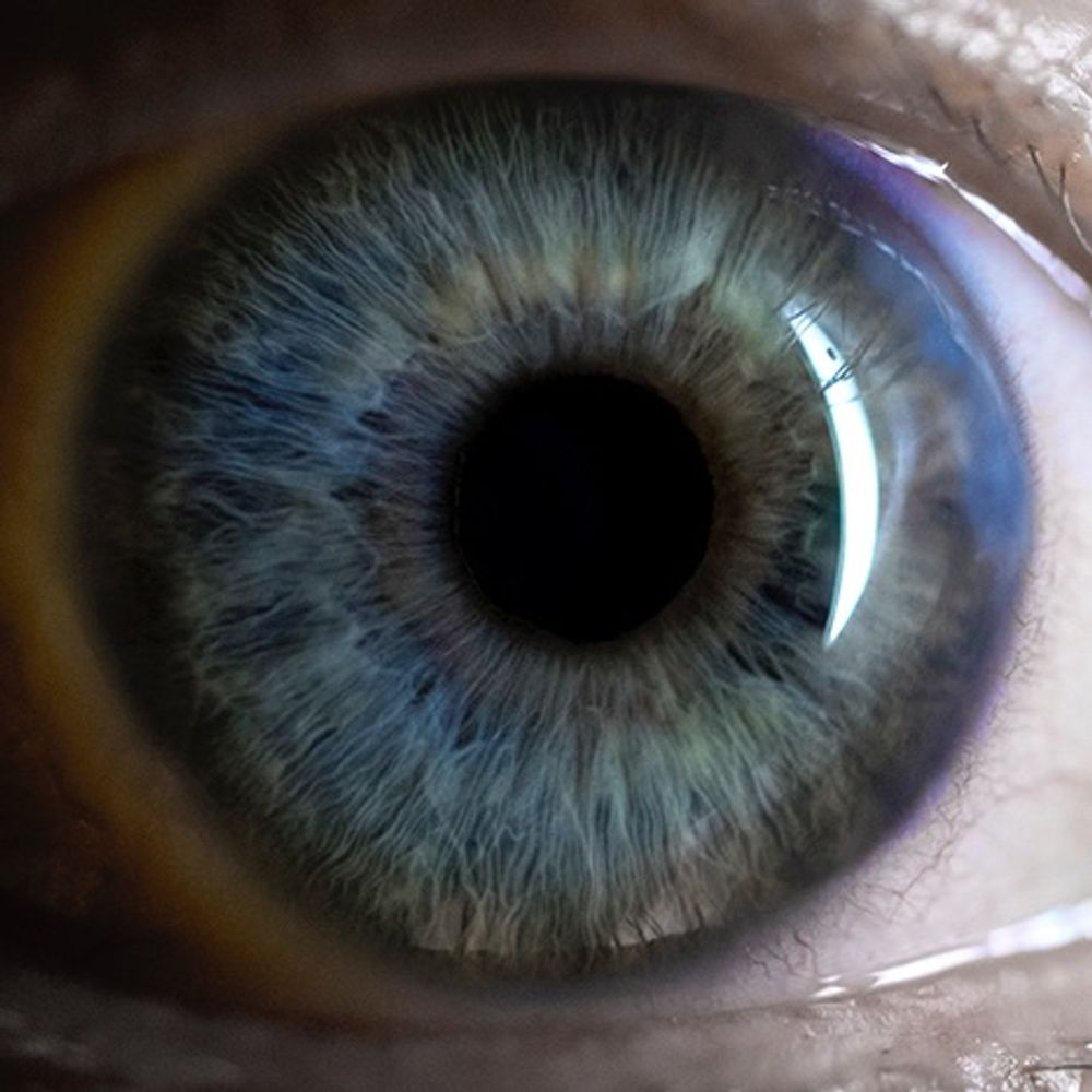
A unique view of a real human eye from the inside, floating in the vitreous.
This video highlights the power of studying a sample in 3D, by immersing the viewer inside the entire eye of a 40yo donor.
24.06.2024 04:52 — 👍 0 🔁 0 💬 0 📌 0
Macular telangiectasia Type 2 (MacTel) is a progressive disease that affects the quality of life by impairing both distant and near vision.
#Retina #Retinopathy #Mac_Tel #V_ophthalmology #VOPHA #Optometry #eyeacuity #ophthotwitter @mazhry @eyeacuity
31.05.2024 04:10 — 👍 0 🔁 0 💬 0 📌 0

Hey #ophthotwitter
We did it! 50 uploads strong on YouTube!
A big thanks to our viewers for helping us reach this milestone.
Subscribe for more eye health content!
#ophthalmology #VOPHA #eyehealth
@mazhry @eyeacuity @MAbdullahMazhry @Laiba68539150
27.05.2024 06:00 — 👍 0 🔁 0 💬 0 📌 0
Greatly valuable information about #CSCR.
#OCT #Retina #choroid #vitreous #Retinopathy #AntiVEGF #V_ophthalmology #VOPHA #Ophthalmology #AAO #Optometry #eyehealth #ophthotwitter
@mazhry @eyeacuity @MAbdullahMazhry @Laiba68539150
15.05.2024 03:42 — 👍 0 🔁 0 💬 0 📌 0
Laser refractive, lens and glaucoma surgeon. Educator, entrepreneur and grew up in Singapore. Love the big smoke in London. Based at St Thomas’ Hospital, Westminster and Wimpole Street. Follow me on LinkedIn and Instagram for practice updates.
Eye surgeon obsessed with futuristic technology and lasers and their use in conquering blindness and rejuvenating sight. Founder of @Centreforsight UK
Working to make healthcare more human.
Speaker. Podcaster. Writer. Advocate.
SCA lay responder.
Co-survivor of cancer (2x), sudden cardiac arrest and medical training.
Hypermobile 🦓
Married to that funny guy.
glaucomflecken.com
@medsky.social
Work: Academia/Ophthalmology - IRD and Retina Surgeon
Life: Reading, games, travel, 🏳️🌈
The Association for Research in Vision and Ophthalmology (ARVO) is an international eye and vision research organization with more than 11,000 members worldwide.
ماہرِ چشم اور سخن ور
آنکھیں روشن دل منور
Mazhry, a renowned Ophthalmologist and Urdu poet, blends the precision of medicine with the beauty of poetry. Based in Lahore, Pakistan, practices ophthalmology and writes diverse Urdu poetry, captivating readers.
Academic / Paediatric Ophthalmologist
Epidemiology, Eyes/Vision, Imaging, Data science, Health equity.
Huge nerd.
Not necessarily in that order.
Both kinds of doctor.
https://www.ted.com/talks/dr_lola_solebo_through_the_eyes_of_a_child?subtitle=en
Ophthalmologist. Comedian. Speaker. Jonathan.
A veterinary ophthalmologist @ucdavis, 👁 scientist, & mom looking to make the 🌎 clearer for people & animals. Co-director of CMSTP T32 for DVMs getting their PhD. Views are my own. 🐴🦜🥰
https://covsl.vetmed.ucdavis.edu/
Ophthalmology-related education.
Optometry’s trusted advocate for the advancement of clinical expertise since 1891. www.reviewofoptometry.com
Ophthalmologist | Senior Research Fellow, UCL | http://windowsofthesoul.art #oculomics All views my own
Inspiring Excellence in Eye Care.
LeadNurse4Research @Moorfields
Prof. of Ophth Health & Care @UCLeye @NIHRresearch Programme Director Senior Research Leaders
@GlaucomaUK trustee
#NIHRSRL #NIHR70at70
🇹🇹
Professor, eEF1A2/neurological disorders. Mostly talks about research, EDI (tries hard to be a good ally) but sometimes veers off into crafts and photos of Scotland. She/her, views own.
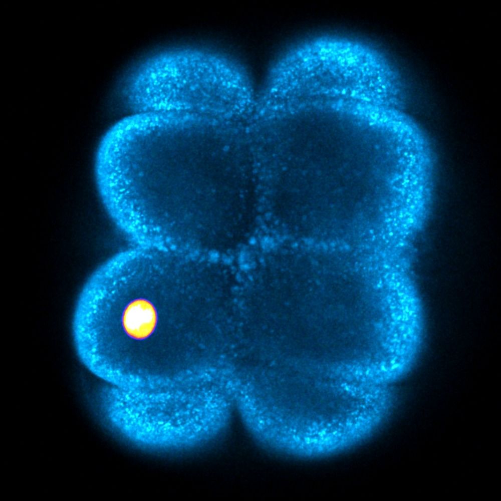
Physics of Embryonic Self-Organization and Morphogenesis. Tweets by Otger Campàs (Professor, Chair of Tissue Dynamics and Director
at the Physics of Life Excellence Cluster of TU Dresden)
At the back of your eyes.
https://webvision.med.utah.edu
Research Fellow at the International Centre for Eye Health at LSHTM
Now not posting here instead of not posting on Twitter.