Sure thing, you’re added!
11.02.2025 09:48 — 👍 1 🔁 0 💬 0 📌 0
Already have you in Cathy 😉
08.02.2025 10:11 — 👍 2 🔁 0 💬 0 📌 0
You’re in my friend 👍🏻
08.02.2025 08:16 — 👍 1 🔁 0 💬 0 📌 0
Sure, you’re in! 👍🏻
15.01.2025 10:04 — 👍 1 🔁 0 💬 0 📌 0
Absolutely, you’re in! 👍🏻
12.01.2025 14:14 — 👍 1 🔁 0 💬 0 📌 0
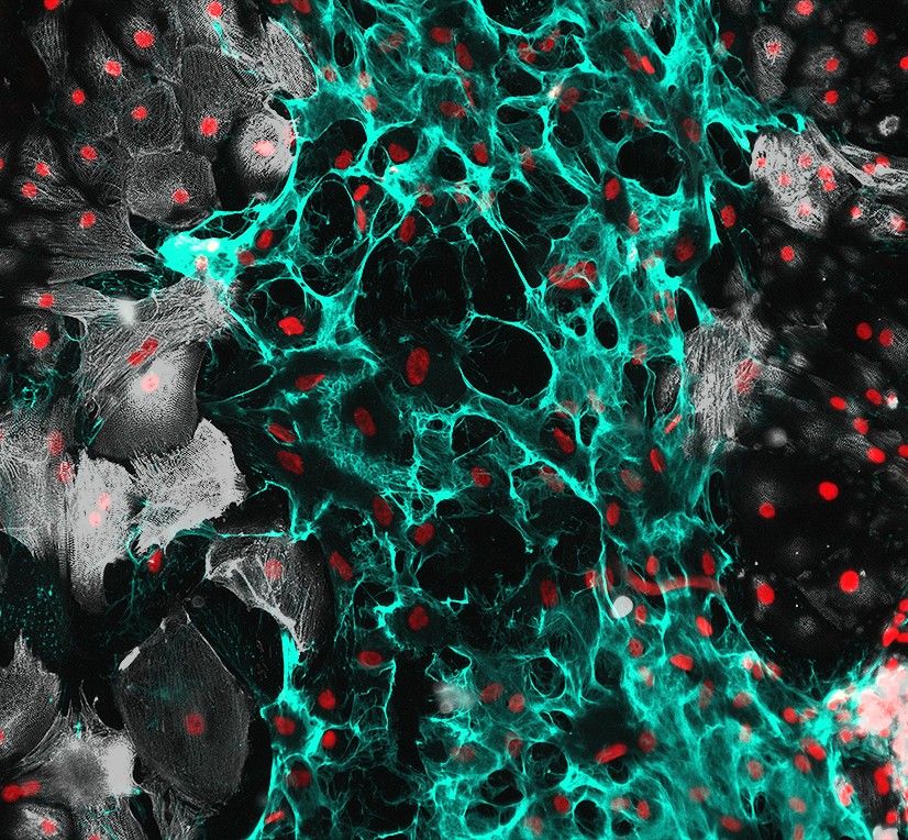
Cardiac scratch wound assay to show the role of ECM in wound healing. HiPSC Cardiomyocytes in white on the boarders with a rich ECM scar tissues (cyan) through the middle with a simple nuclear stain (red) highlighting all cells.
How to fix a broken #heart 💔->❤️
#Proteoglycan rich #ECMatrix scar tissue is laid by migratory cells after a cardiac scratch wound with #hiPSC #Cardiomyocytes.
Happy #Fluorescencefriday #BlueSky & #CardioSky!
@ablanchasensio.bsky.social 👀
#Science 💥🧪 #Microscopy 🔬
13.12.2024 15:22 — 👍 61 🔁 12 💬 0 📌 0
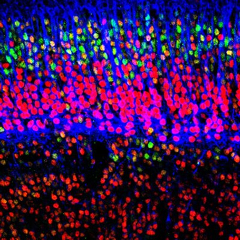
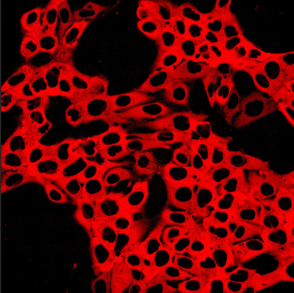
DNA during cell death and cell division.
#Science #Biology #CellBiology #microscopy
11.12.2024 14:20 — 👍 35 🔁 8 💬 0 📌 0
Sure, done!
09.12.2024 09:24 — 👍 0 🔁 0 💬 0 📌 0
👍🏻
08.12.2024 22:46 — 👍 0 🔁 0 💬 0 📌 0
Sure thing, you’re in!
08.12.2024 11:53 — 👍 1 🔁 0 💬 0 📌 0
Added! 👍🏻🧪
08.12.2024 09:05 — 👍 0 🔁 0 💬 0 📌 0
Sure, done! 👍🏻
08.12.2024 07:11 — 👍 0 🔁 0 💬 1 📌 0
Hi James, would love to be added thanks!
08.12.2024 07:08 — 👍 0 🔁 0 💬 0 📌 0
Genetically-modified hiPSCs expressing the genetically-encoded calcium indicator jRCaMP1b with a nuclear export signal (NES). This reporter is used to track calcium dynamics in live cells. It combines a red fluorescent protein with calmodulin and a calcium-binding domain, changing fluorescence intensity upon calcium binding. Its red-shifted emission is ideal for imaging in complex tissues or dual-color experiments, making it valuable for studying calcium-mediated processes.
A single copy of this reporter was integrated using the STRAIGHT-IN platform at the AAVS1 locus. See below!
https://doi.org/10.1016/j.crmeth.2022.100300
https://doi.org/10.1101/2024.10.17.616637
Here is my 2nd #FluorescenceFriday post! 🧪 #hiPSCs expressing jRCaMP1b, a genetically-encoded Ca2+ indicator 👻. This reporter allows us to record intracellular Ca2+ in hiPSC-derived #cardiomyocytes and cardioids (videos in the comments) #microscopy 🔬 #Science #Biology @benjaminbjohnson.bsky.social
06.12.2024 14:59 — 👍 27 🔁 3 💬 2 📌 0
There are so many #morphogenesis researchers here!
My first starter pack is full!
Here is Part 2: go.bsky.app/K6YYKeC
#devbio #cellbio #science
Tell me if I should add you but try to make sure, that you're not already in Part 1.
I cannot cross-reference everyone 😅
Part 1: go.bsky.app/RzDD9or
27.11.2024 18:00 — 👍 48 🔁 21 💬 20 📌 3
Hi, could I be added to your pack thanks!
06.12.2024 15:56 — 👍 2 🔁 0 💬 0 📌 0
Hey Jeff, would love to be added to your pack thanks!
06.12.2024 15:55 — 👍 1 🔁 0 💬 0 📌 0
Hey, would love to be added! I work on hPSC cardiac systems and disease models ❤️
06.12.2024 15:52 — 👍 1 🔁 0 💬 0 📌 0
Hey, would love to be added thanks! 😀
06.12.2024 15:50 — 👍 0 🔁 0 💬 1 📌 0
Hey, would love to be added to your pack thanks! ❤️
06.12.2024 15:48 — 👍 0 🔁 0 💬 1 📌 0
Hey, can I be added please and thanks! 😀
06.12.2024 15:44 — 👍 1 🔁 0 💬 1 📌 0
Hey! Would love to be added to this pack, thanks! ❤️
06.12.2024 15:41 — 👍 1 🔁 0 💬 0 📌 0
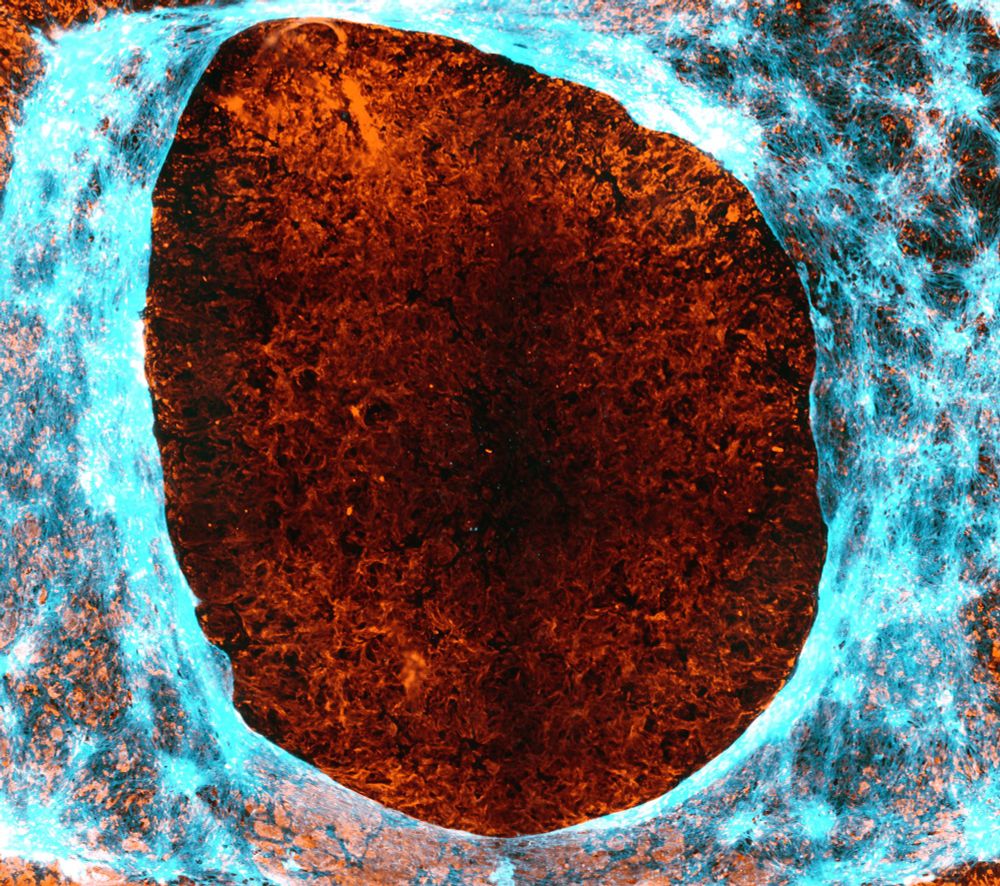
Cyan cellular extracellular matrix fibers are highlighted laying over the top of the orange hyaluronan secreted matrix after a scratch would assay.
Dedicating this #Fluorescencefriday to the #ECMatrix lovers. Was trialing some ECM staining methods on a scratch wound and love the HABP staining looking almost like flames 🔥🧪🔬
Lets see what you got, @ablanchasensio.bsky.social 😉
06.12.2024 14:27 — 👍 20 🔁 2 💬 0 📌 0
Definitely, you’re in!
06.12.2024 07:03 — 👍 1 🔁 0 💬 1 📌 0
Absolutely, you’re in! 👍🏻
05.12.2024 20:32 — 👍 2 🔁 0 💬 0 📌 0
Done!
05.12.2024 20:19 — 👍 0 🔁 0 💬 1 📌 0
Created a starter pack focusing on disease modeling in a dish using stem cells! 🧪🔬
Please (self)nominate anyone you think should be added to the list and repost so it finds the right people ❤️
hPSC Disease Modeling: go.bsky.app/EpWsxNG
For a good general stem cell pack go here: go.bsky.app/UTW4d7b
05.12.2024 15:53 — 👍 39 🔁 16 💬 14 📌 1
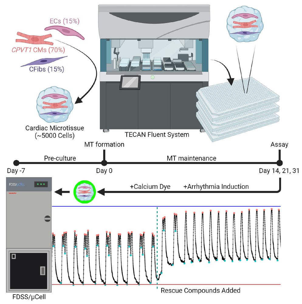
Graphical abstract depicts automated setup to generate 3D cardiac mini tissues consisting of 3 cell types seeded into 384 well plates. The cardiac tissues are monitored using calcium traces to detect arrhythmias and subsequent rescue after compound addition.
🔥Super excited to drop our recent preprint🔥
biorxiv.org/cgi/content/...
Here, we automated the generation of 3D Cardiac Microtissues and performed a small high-throughput screen to identify several novel anti-arrhythmic compounds for CPVT1 🧪❤️
#Cardiosky #Science #hiPSC #Arrhythmia #Stemcell
04.12.2024 15:13 — 👍 13 🔁 3 💬 1 📌 0
Science! 🧪
29.11.2024 14:36 — 👍 1 🔁 0 💬 0 📌 0
Hey #BlueSky! Here is my first #FluorescenceFriday post!
This cardiac organoid was derived from #hiPSC and imaged using light sheet #microscopy 🔬.
🔵 Cardiomyocytes
🔴 Endothelial cells
🟡 Fibroblasts
More coming next week! 🧪
#Science #Biology #CellBiology #SciArt
@benjaminbjohnson.bsky.social
29.11.2024 14:05 — 👍 51 🔁 5 💬 4 📌 0
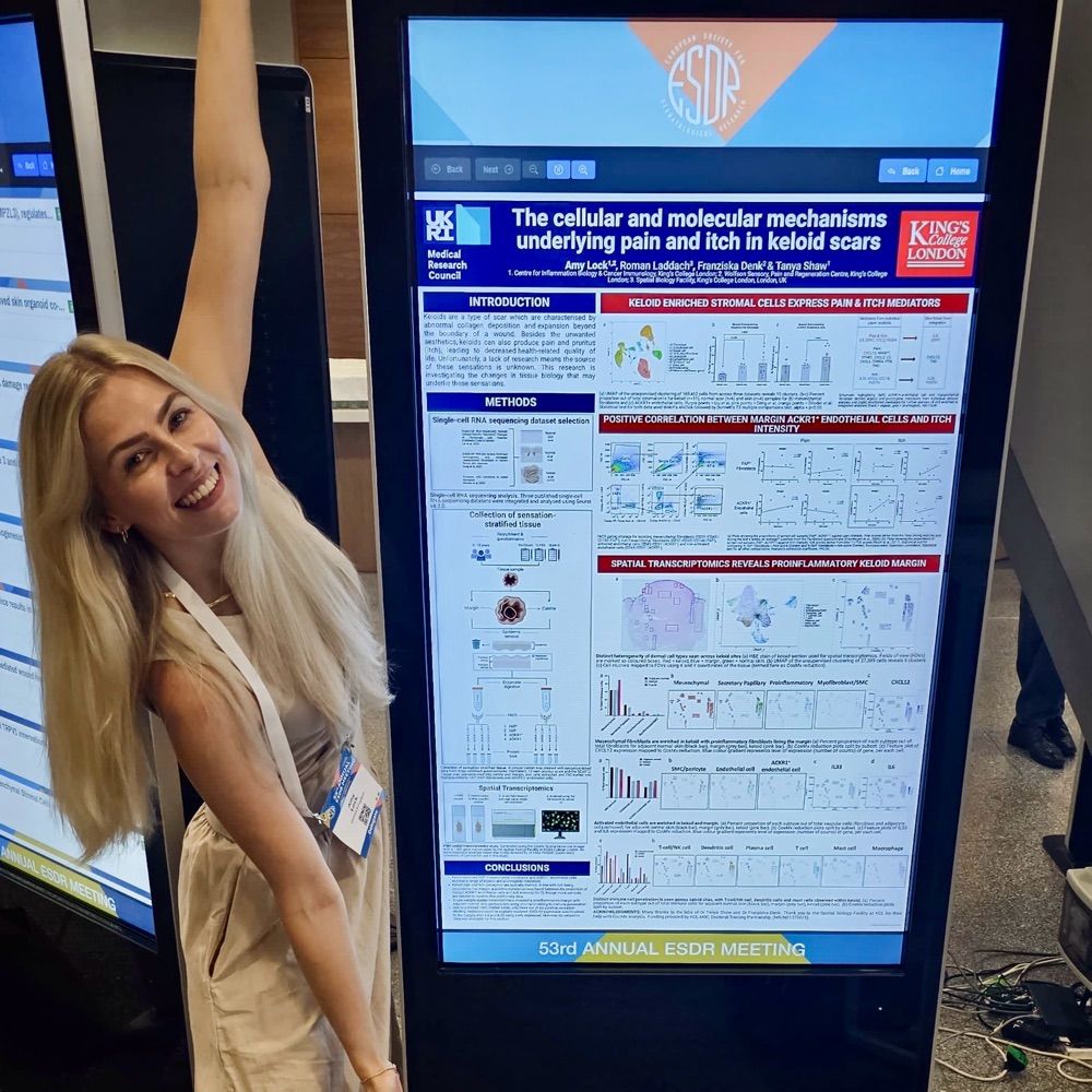
Postdoctoral researcher in Denk lab and Shaw lab. Researching aberrant sensations in keloid scars.
stromal cells | single cell sequencing | iPSC sensory neurons | clinical & translational | pain & itch
Post-doctoral researcher working in stem cell, RNA and cardiovascular research at UEA Medical School
Developmental Biology & Stem Cell research network
@ Norwich Research Park, U.K.
https://www.norwichdevbiostem.org.uk/
Academy Research Fellow | Mitochondrial Biology and Metabolism | Stem Cell Research | Neurodegenerative disorders Models | Passionate about history, sci-fi literature, and miniature painting. Helsinki, Finland. 🇪🇸 🇫🇮
Sr. Scientist at Cardiac Organoid start-up HeartBeat.bio, former PhD Christine Mummery lab.
Cardiac organoid disease models | High-throughput screening | Pluripotent Stem Cells | Omics
3D iPSC models and CRISPR to investigate mechanisms of neurodegenerative and neurovascular disorders | Professor of Neurobiology at LMU Munich | https://isd-research.de/paquetlab
The Young-Pearse lab focuses on the identification of the mechanistic causes of neurodegenerative and neurodevelopmental disorders. Account run by lab members.
Find us at: youngpearselab.bwh.harvard.edu
An open-access @natureportfolio.bsky.social journal publishing high quality primary research, reviews and commentary in all areas of the biological sciences since 2018.
https://www.nature.com/commsbio/
An open access @NaturePortfolio journal publishing high-quality primary research articles, reviews and commentary in all areas of the chemical sciences.
nature.com/commschem/
An open access @natureportfolio.bsky.social journal publishing high quality primary research, reviews, and commentary in Earth, environmental, and planetary science.
nature.com/commsenv/
The multi-disciplinary engineering journal from the Nature Portfolio. Fully open access. You can read all our content here for free: https://www.nature.com/commseng/
An open access journal from Nature Portfolio publishing important advances in all areas of materials science.
A selective open access journal from @NaturePortfolio publishing research, reviews & commentary across clinical, translational & public health research fields. nature.com/commsmed
Communications Physics is an open access journal from Nature Portfolio publishing high-quality research, reviews and commentary in all areas of the physical sciences.
https://www.nature.com/commsphys/
Communications Psychology is a selective, peer reviewed, open access journal in the @natureportfolio.bsky.social, publishing high-quality research, reviews and commentary across psychology.
https://www.nature.com/commspsychol/
A Nature journal dedicated to presenting the very best research across the disciplines of astronomy, astrophysics, cosmology and planetary science.📡
www.nature.com/natastron
Publishing the latest advances across all areas of cancer research and oncology. Part of @natureportfolio.nature.com
🌐 www.nature.com/natcancer/
📍New York, London, Berlin and Heidelberg
Nature Catalysis publishes high quality work across all areas of catalysis, including both fundamental and applied studies.
https://www.nature.com/natcatal/
A journal dedicated to publishing the latest advances across all areas of cell biology. Part of @natureportfolio.nature.com
nature.com/ncb/index.html