
Fauja Singh, world's oldest matrathon runner
A Super Ager, Fauja Singh, ran his 1st marathon at age 89, and 8 more after that through age 101.
How does he die at age 114 years?
Hit by a car.
apnews.com/article/olde...

@ucpath.bsky.social
Veterinary pathologist, conservation advocate, wildlife-disease scientist, outdoorsman. And I love parasites, but especially the metazoans! Scientific, one-health and naturalist content.
Fauja Singh, world's oldest matrathon runner
A Super Ager, Fauja Singh, ran his 1st marathon at age 89, and 8 more after that through age 101.
How does he die at age 114 years?
Hit by a car.
apnews.com/article/olde...
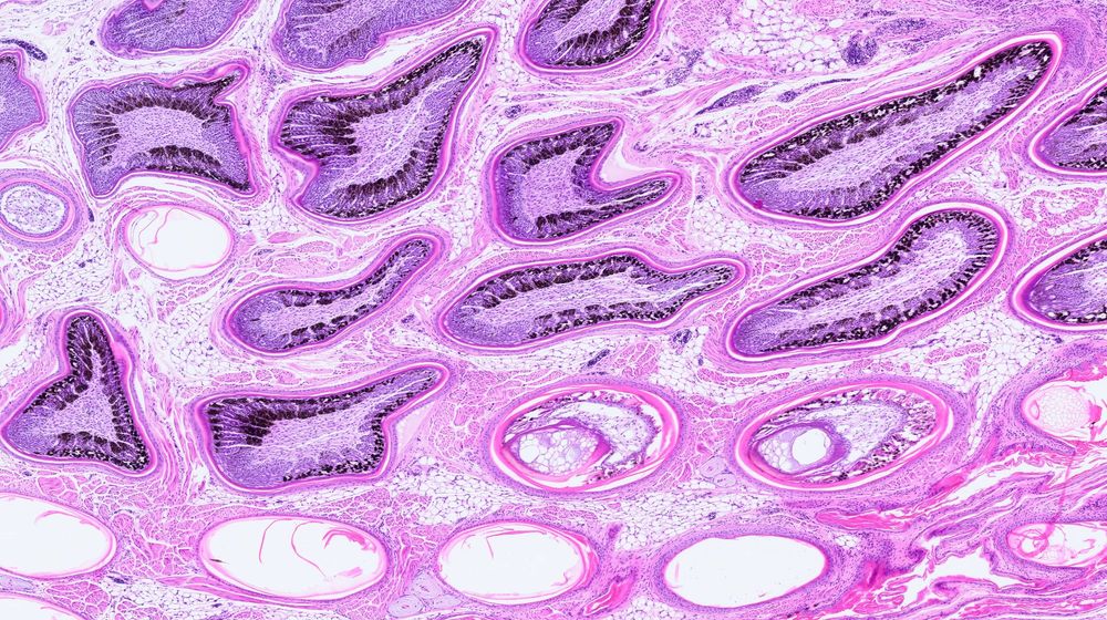
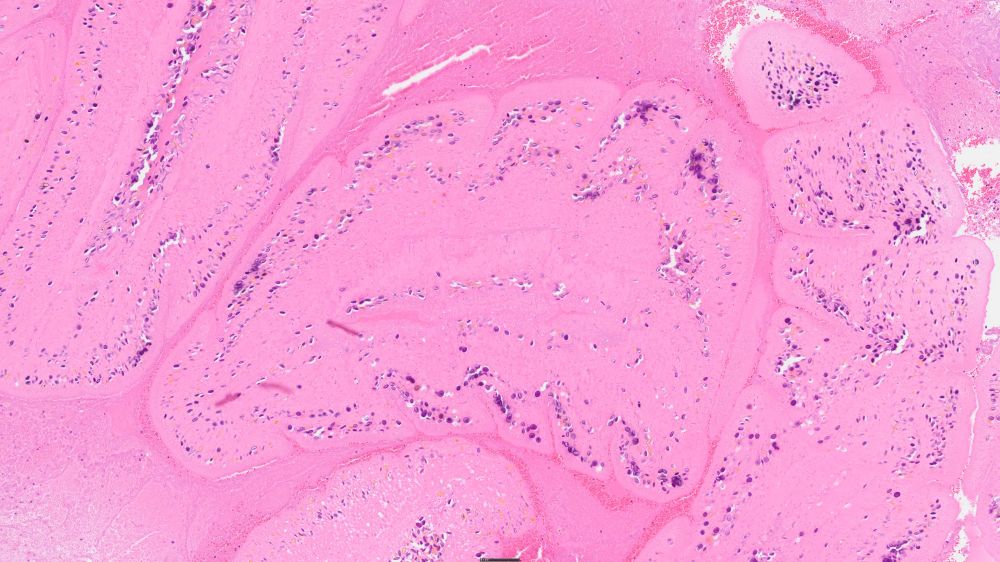
Beautiful representation of feather follicles from a chickadee. A happy accident from the histology lab. They are not supposed to be sectioned obliquely but it makes them look pretty interesting in an abstract sort of way. 🪶
16.07.2025 21:42 — 👍 9 🔁 0 💬 0 📌 0I wish it were that easy to stop my sneezing.
02.07.2025 16:52 — 👍 1 🔁 0 💬 1 📌 0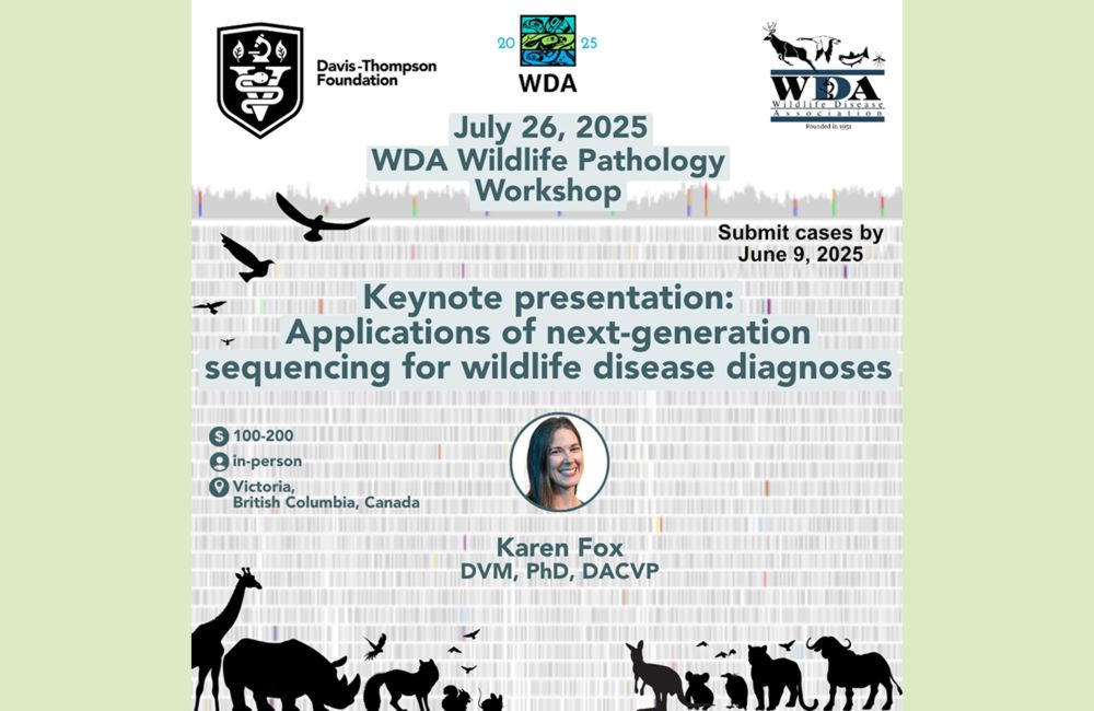
Submit your case for the Wildlife Pathology Workshop at #WDA2025
📅 Deadline for slide submission: June 9
🔬 Focus: Gross & histopathology of wildlife cases
📍 Held during the WDA Annual Meeting
#VeterinaryPathology #WildlifeHealth #OneHealth
wda2025.com/event/wda-an...
It reminded me of the $6.2 million banana with duct tape.
12.04.2025 16:19 — 👍 2 🔁 0 💬 0 📌 0
With 1,000 dairy herds affected by avian influenza and the disease regularly changing, the absence of this program is going to hurt.
www.reuters.com/business/hea...
It's good to see these coming back. There has been a huge difference in the population since I returned to California 13 years ago.
03.04.2025 23:40 — 👍 2 🔁 0 💬 1 📌 0
A sea lion with domoic acid poisoning experiencing involuntary muscle spasms. This is a female sea lion lying on her side on a beach. Her muzzle and left flipper pointing skyward. She has domoic acid poisoning and was experiencing involuntary muscle spasms. Credit: Channel Islands Marine & Wildlife Institute
California is no stranger to domoic acid, but this year it's much worse much earlier.
Stranded pinnipeds often exhibit listlessness, head bobbing, disorientation and seizures. Mortality rates are estimated to be ~25%
www.fisheries.noaa.gov/feature-stor...
#VetMed #MarineMammal #VetPath
Very cool. I saw my first fisher this past weekend. But, it was on the necropsy table. Most people have a life list...
31.03.2025 20:28 — 👍 1 🔁 0 💬 0 📌 0Well, shit.
This is going to cost us a lot more than the money saved. For the price of 38 Abrams tanks, #FAO has gotten a hell of a lot done with support from the U.S.
kbhbradio.com/funding-term...
#VetMed #AnimalHealth #OneHealth
This seems a bit obvious, but it's a very cool illustration of the idea. I tried to figure out where it was first published, but all I found were many social media posts with the image. Guilty!
31.03.2025 14:25 — 👍 0 🔁 0 💬 1 📌 0
graphic of branching lifelines, with the path taken traced through it. past paths no longer in play are greyed out, possible future paths spread out like a network of roots in front
The future is a web of branching paths, and each choice opens up new possibilities as it closes others. This is as true for a civilization as it is for an individual. The more our collective intelligence can inform each choice, the better our chances of finding paths through to a beautiful future.
31.03.2025 06:22 — 👍 9181 🔁 1377 💬 342 📌 143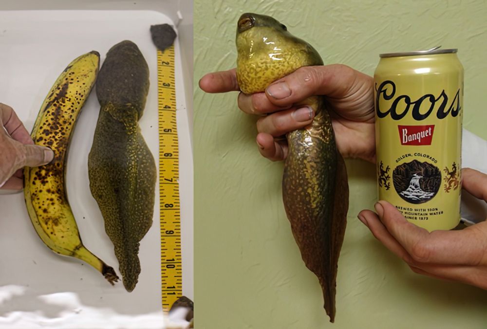
Photos of a giant bullfrog tadpole. the panel on the left compares it so a similarly sized banana, and a tape measure showing that it is nearly 11 inches long. The right panel compares it to a beer can. It is not the first time I have seen someone use a beer can or a beer bottle as a size reference. I guess you use what you've got. And they are a constant size.
Not a new article, but it's worth posting again just in case you missed it. Bullfrogs are normally tadpoles for 2 or 3 years. Something disrupted this one's metamorphosis so it just kept on growing! I love the references for scale!
#frogs #wildlife
www.americanscientist.org/blog/from-th...

If only we had a vaccine for ignorance. But, wait! We do!!
Or, at least we did. Unfortunately, critical thinking and education, it would seem, are passé.
www.nbcnews.com/health/healt...
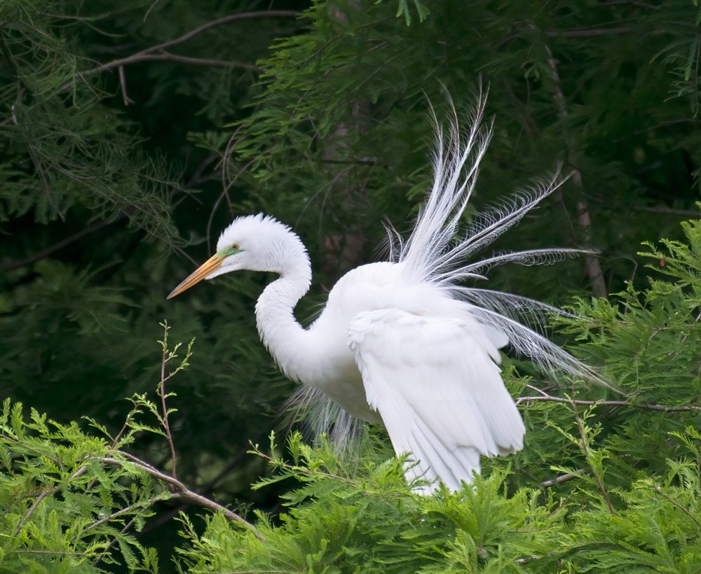
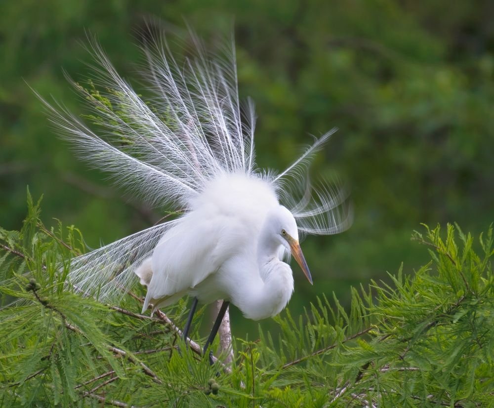
This is a profile of a common egret in full display, sitting in they top of a cypress tree. The nuptial plumes are Lacey feathers protruding from the body. The bird also has a green cere, that is a result of its breeding status.
An exhibitionist egret on display. These courtship threads beat millinery any day. Populations of common #egrets (and other spp.) were once decimated so we could put their nuptial plumes in women’s hats. Goshen Swamp, Liberty Co, Georgia.
#birds #wildlife #NaturePhotography #Photography
Kidneys are an amazing organ! And the lesions always make sense to me. I can wrap my head around renal lesions in a way that I never could quite master with repro pathology.
19.03.2025 23:49 — 👍 1 🔁 0 💬 0 📌 0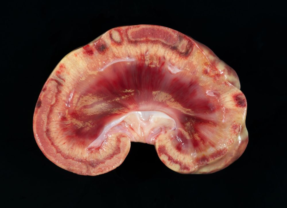
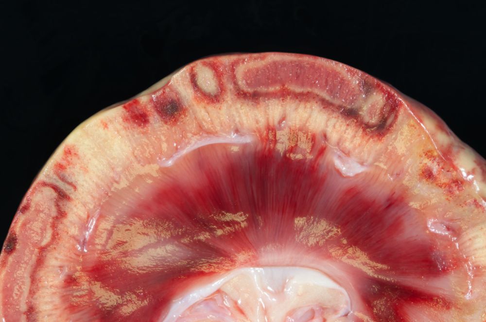
This is the cut surface of a kidney that has been sectioned longitudinally. Six poorly demarcated, dark-red, wedge-shaped foci extend from the corticomedullary margin to the pelvis and are 4 to 7 mm wide at the corticomedullary junction. One, which corresponds to a cranial cortical depression, is itself slightly depressed on cut surface (infarct).The cortex of the left kidney has five, focal to focally extensive discolored foci that are restricted to the superficial one-half of the cortex. The smallest of these are rectangular, dark red foci in which each edge of the rectangular focus is approximately 5 mm. The largest of the superficial cortical foci consists of a 6 cm long band in the outer one-half of the cortex. All of the larger foci have a pink core with a ground glass surface (necrosis) surrounded by a 1-mm-thick yellow margin that is in turn surrounded by a 1-mm-thick, red outer margin of tissue. Close inspection reveals congested glomeruli in the less affected cortex, but these are not visible in the necrotic regions.
A macroscopic image of the canine kidney previously described. Less affected regions are uncharacteristically yellow. There are scattered aminar, supericial-cortical lesions. The smallest are dark red. The largest have a pink core with a ground glass surface (necrosis) surrounded by a 1-mm-thick yellow margin that is in turn surrounded by a 1-mm-thick, red outer margin of tissue. If you look closely you can see pin-point red structures scattered throughout the cortex that correspond to glomeruli.
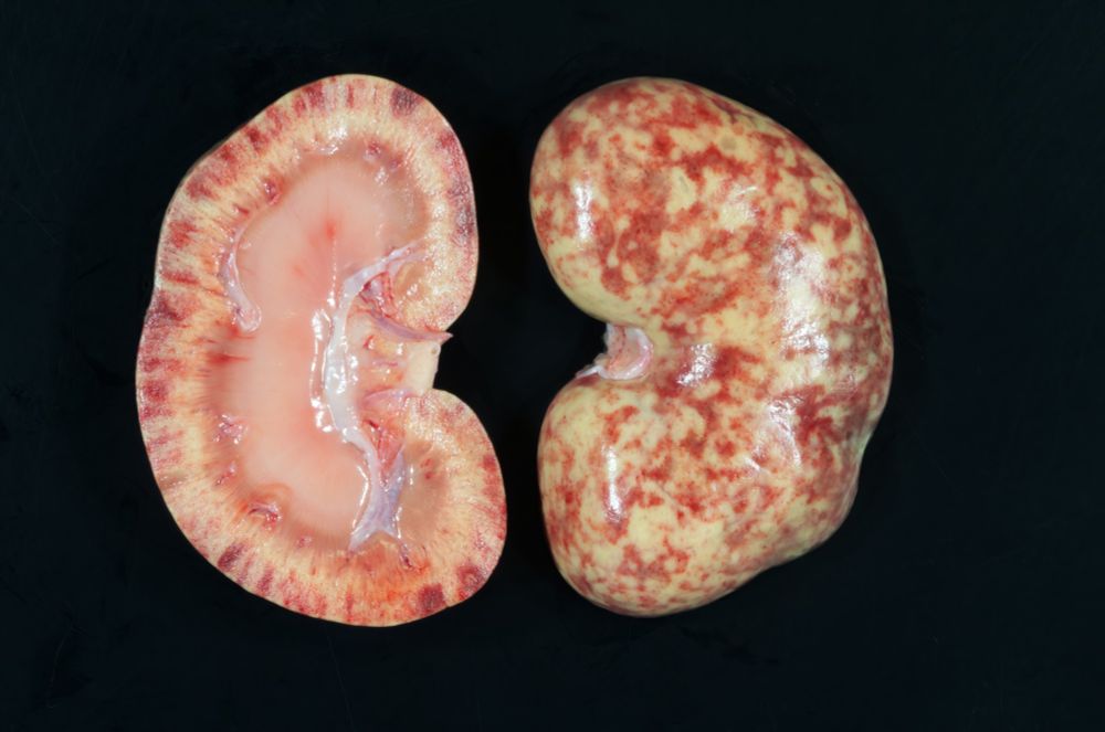
The image depicts the right kidney from this case that has been sectioned longitudinally through the hilum. The section on the left shows the cut surfaces. The one on the right shows the capsular surface with the capsule removed. The capsular surface is speckled red and yellow with hundreds of widely scattered, pinpoint, red foci that are only visible with very close inspection (suspect congested or hemorrhagic glomeruli). The larger red foci account for half of the surface of the right kidney and they extend into the outer one-half of the cortex on cut surfaces. The kidney is otherwise pale.
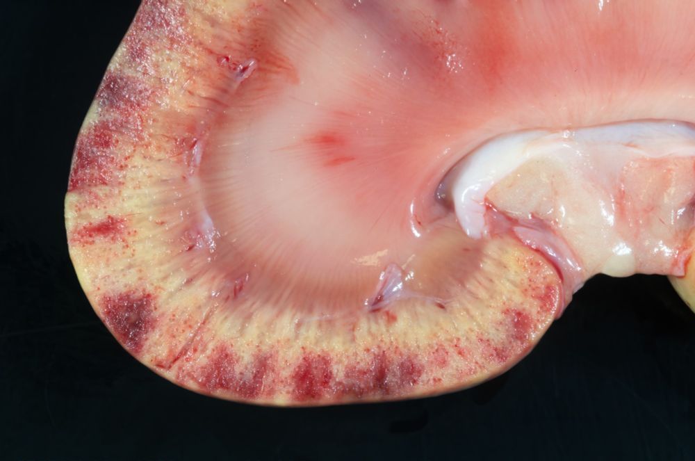
This is a macroscopic photo of the right kidney. On cut surface the red foci are mostly limited to the superficial one-half of the cortex. Pinpoint red dots are clearly visible here, and they likely correspond to discolored glomeruli. The remainder of the cortex is yellow and it should be reddish brown.
These are some crazy kidneys! You don't have to be a pathologist to think these lesions are beautiful!
I have suspicions as to pathogenesis, but histo will tell.
9-yo dog. Clinicians susp granulomatous osteomyelitis of lumbar v but no gross lesions were evident.
#pathsky #vetpath #RenalPath
A common egret displays its plumes in the top of a cypress tree. The bird is holding its neck in a semicurl and the plumes are spread behind it like a snow white peacock. The photograph resulted in the whites being a little blown out in the birds back. Hard to get everything balanced with these white birds in a dark swamp.
It's that time of year again! The #egrets are getting frisky. My parents are lucky enough to have a rookery in their backyard.
Common egret, Liberty Co, #Georgia
#birds #nature #wildlife #NaturePhotography #Nikon #swamp
Essential thrombocythemia I presume. I had to refresh my memory on this. Schalm's has a nice section on it, but all the acronyms drive me a bit nuts in that book. I get frustrated when I have to go back to the beginning to constantly remind myself what they mean by PV, CMPV, CALR, IDA, ET, etc!
19.03.2025 15:43 — 👍 1 🔁 0 💬 0 📌 0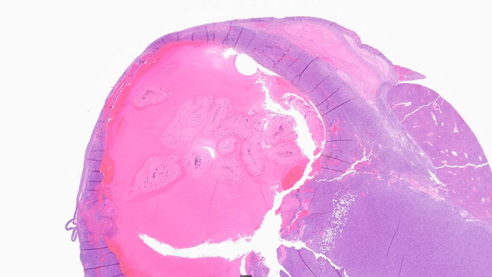
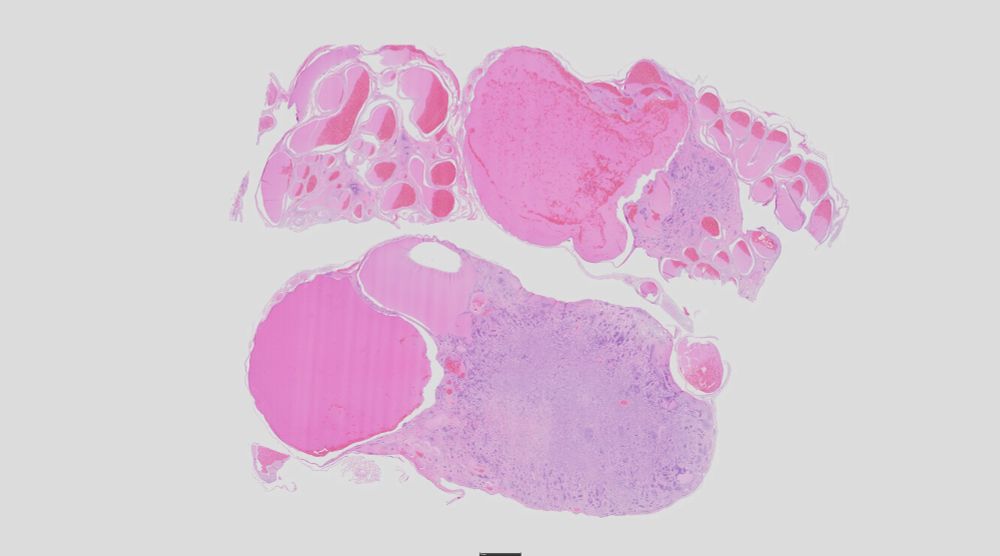
This is a subgross image of liver with a large tumor containing a blood filled cavity that has multiple sections of tapeworm larvae. A bit of the hepatic parenchyma is on the upper right. H&E-stained section.
Here are cross sections of the degenerate tapeworm larvae from the previous image. They lack detail but the calcareous corpuscles are still visible. These appear to be strobilocerci. As I understand it, this tapeworm initially forms cysticerci in the rat which then metamorphose into strobilocerci. H&E-stained section.
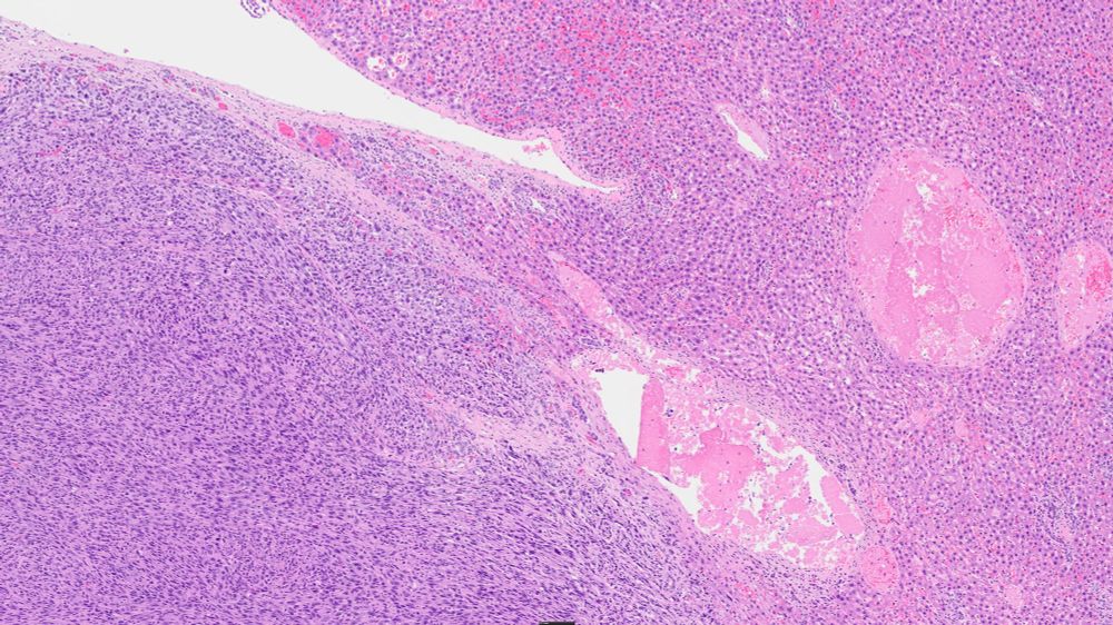
This image demonstrates the fibrosarcoma on the right and the interface with the extant hepatic parenchyma on the left. The neoplasm is well demarcated in this section. In others it was more directly invasive and widespread metastases were all through the abdomen. This seemed to be a case of true sarcomatosis. Implantation of bits of neoplasia directly on the serosal surfaces of the abdomen, leading to additional tumors.
A high magnification image of this neoplasm. Neoplastic cells have significant variability in size. They are generally oval to round. Mitotic figures and apoptotic cells are occasionally seen.
Here's a case you don’t see every day! A hepatic sarcoma in a rat caused by tapeworm larvae. Taenia taeniaformis infestation with chronic inflammation and spindle-cell proliferation paves the way for neoplastic transformation.
Nature is weird sometimes!
#skypath #vetpath #parasites
I should have mentioned that RFK is the other well known cat who had a tapeworm larvae in his brain.
This cat will survive. Luckily, he's not likely to seek a career in politics. That was a pretty big hole in his brain!
#VetPath #Histopathology #Parasites
bsky.app/profile/ucpa...
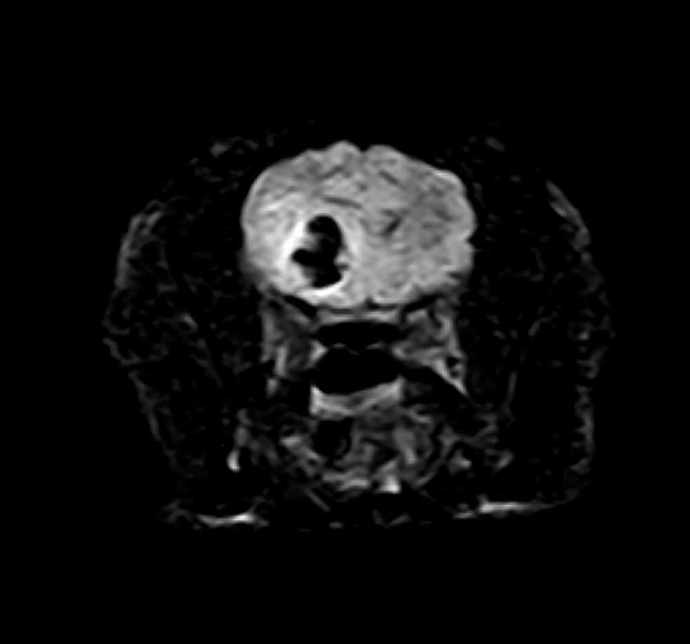
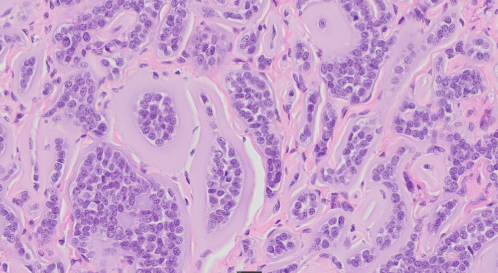
MRI of a cat brain. There is a cystic mass in the right temporal lobe that extended to the frontal lobe.
Subgross section of the biopsy sample from this cat. Multiple scolices are attached to the inside of the cyst visible on MRI. This is consistent with a coenurus. Taenia sp., including Taenia multiceps, Taenia serialis, Taenia brauni, and Taenia glomeratus are capable of producing coenuri. A hydatid cyst could have a similar appearance but a coenurus is more likely given the signalment and location of the patient. PCR and sequence results are pending.
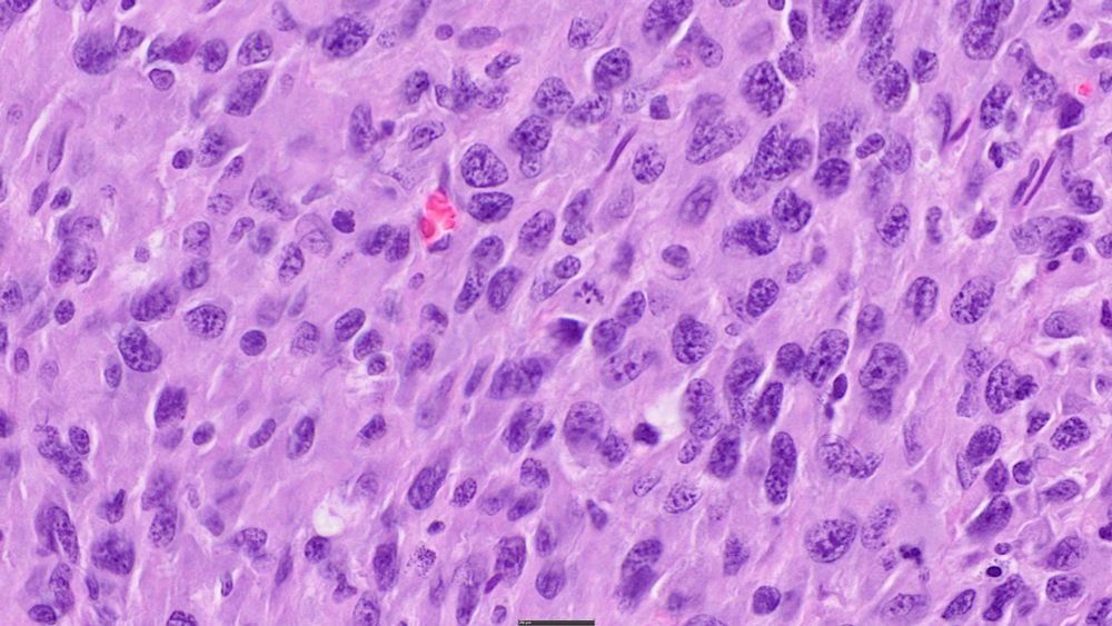
Histopathology slide demonstrating four scolices with hooks and suckers that are variably visible in this section. The presence of multiple scolices in a cysticercoid tapeworm larvae indicates a coenurus or a hydatid cyst. This was most likely a coenurus due to a Taenia sp.
A detail of the scolex from the coenurus of the cat presented. Hooks are evident in the middle of the field with suckers to either side of it. It will have four suckers in total, but the others are out of the plane of section. Calcareous corpuscles are visible in the stroma.
Coolest biopsy of the week! A coenurus from the brain of a cat. This tapeworm larvae has multiple invaginated scolices.
The cat was doing well when discharged 7 days post op.
#VetPath #PathSky #Parasites #Histopathology
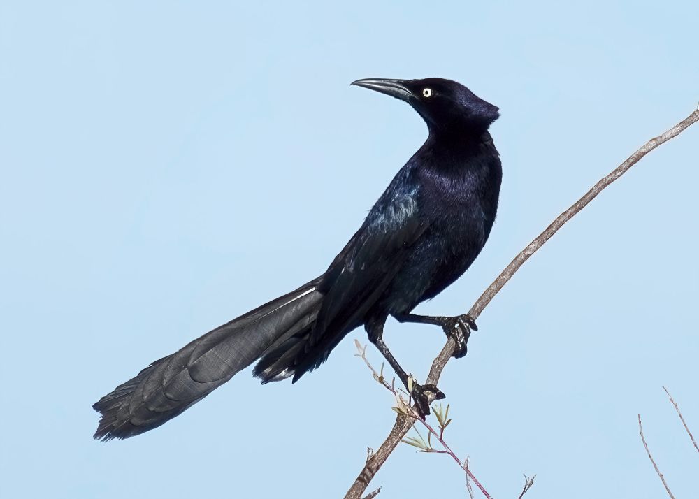
A long, sleek blackbird, a great-tailed grackle (Quiscalus mexicanus) sits perched on a limb looking over his back. The dark irridescent plumage indicates that this is a male. He has a piercing yellow eye, a very long beak and a large tail that is slightly longer than his body.
The great-tailed grackle. My most favorite of all the grackles. Look at that tail! Look at that beak! He is seems pretty proud of the whole package. Woodland, CA.
I wonder how long before they try to change the name to Quiscalus americanus.
#birds #wildlifephotography #naturephotography
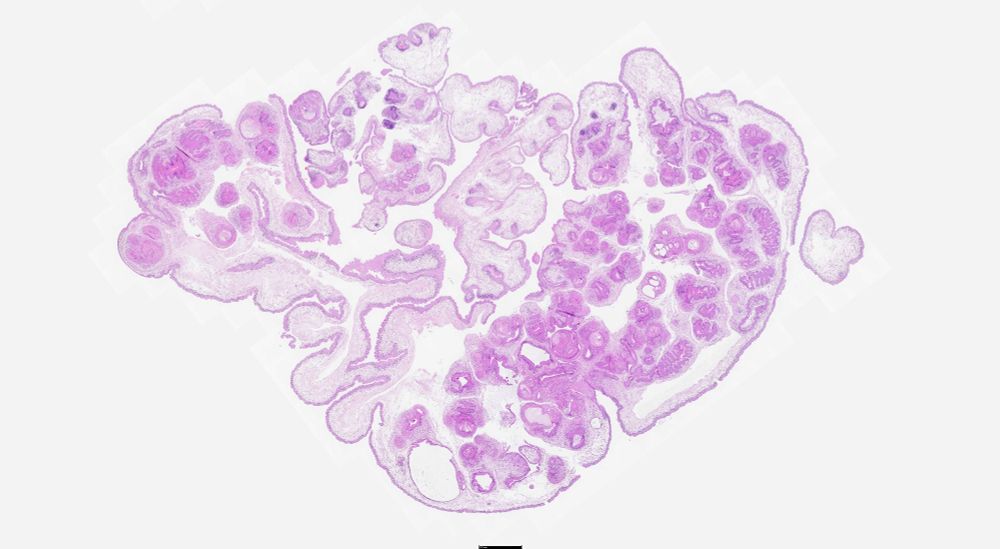
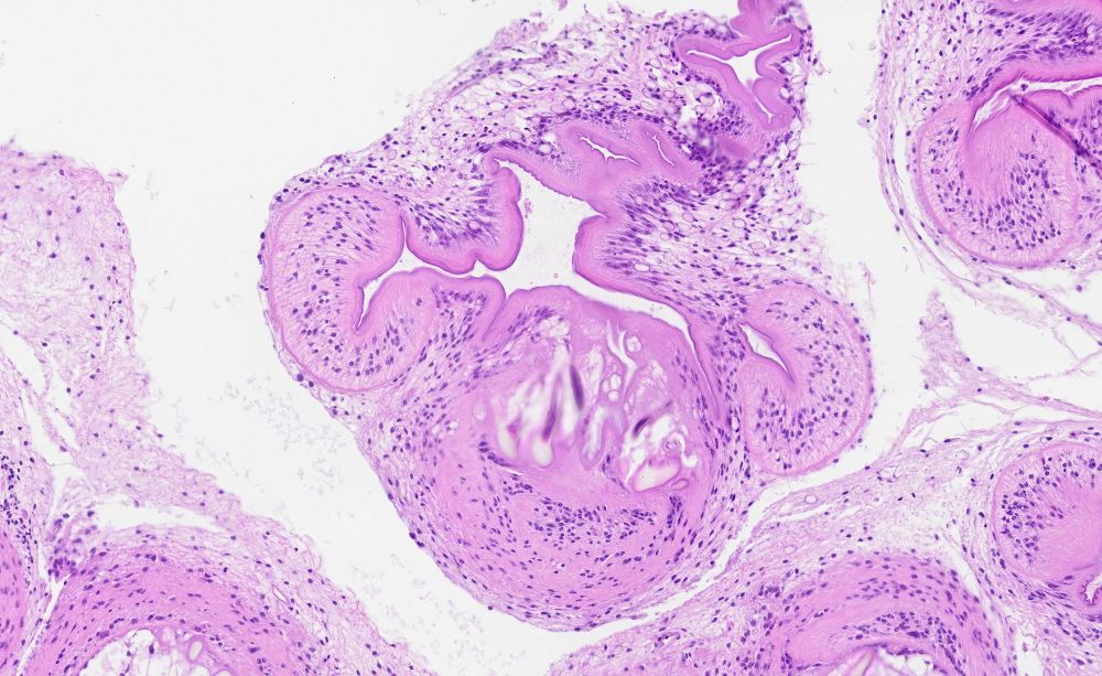
Two sections ostensibly of ovary are present on this microscope slide. The subgross reveals the slightly basophilic masses with tortuous blood vessels adjacent to them.
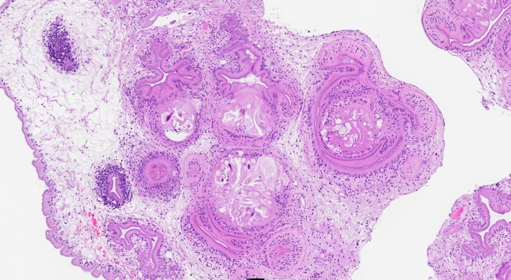
A high magnification view of an ovarian neoplasm from a giant garter snake. The tumor is composed of cords and islands of epithelial cells surrounded by a wide margin of amphophilic material interpreted to be basement membrane material.
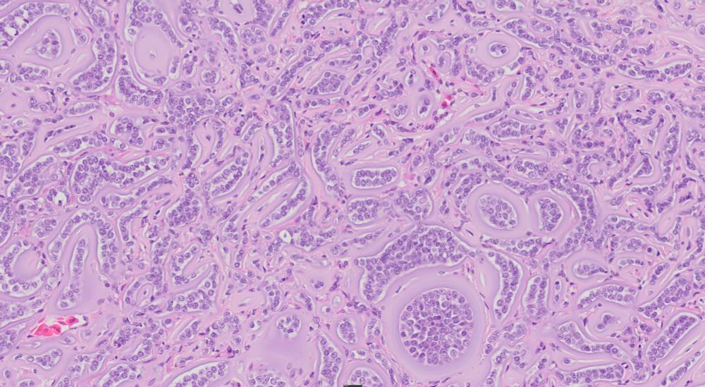
Alternate high magnification view of an ovarian neoplasm from a giant garter snake. The tumor is composed of cords and islands of epithelial cells surrounded by a wide margin of amphophilic material interpreted to be basement membrane material.
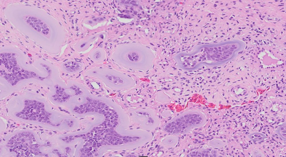
Another high magnification view of the ovarian neoplasm from a giant garter snake. The tumor is composed of cords and islands of epithelial cells surrounded by a wide margin of amphophilic material interpreted to be basement membrane material.
Looking for opinions on this (alleged) ovarian mass from a garter snake. I presume the amphophilic material is redundant basement membrane. Could this be a granulosa cell tumor? We have seen those in snakes (incl garter snakes) but this one is odd.
#pathsky #vetpath #veterinary #tumor #repro
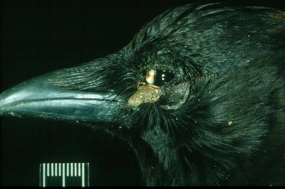
Lateral view of an American crow head. The inferior eyelid has a papillomatous mass that extends from the surface. This was confirmed to be a case of avian pox.
I think it is almost certainly avian pox. Here is one on the eyelid of another crow. I should bring my telephoto lens to try for a better photo of this bird. It has been hanging around for a while now.
18.02.2025 23:15 — 👍 1 🔁 0 💬 0 📌 0What's your diagnosis? American crow with a lesion on the back of the shank. I've seen this bird every day for a week and a half.
#VetPath #AvianDisease #Veterinary #DermPath
Good to know. I was hoping they might be small enough to not cause harm. I wasn't aware they are so well sealed in an airpod. It has been an issue with our dogs. They think they are candy. Probably because they've been in our ears for so long! 😬
05.02.2025 20:48 — 👍 1 🔁 0 💬 0 📌 0I couldn't decide if it was an airpod or a hearing aid. I had a discussion about dogs eating airpods just yesterday. Is the battery large enough to do them serious harm?
05.02.2025 20:34 — 👍 1 🔁 0 💬 1 📌 0
This photo shows a river otter lying on a log on the bank of a river. The otter is very wet and is staring into the camera. There seems to be a scar on his lip, I presume from fighting.
Normally we go to Lake Solano (Yolo Co, California) to see the migrator waterfowl, especially the buffleheads and goldeneye. This day the #RiverOtters were putting on more of a show.
#mammals #WildlifePhotography #Nikon #wildlife
Here is the article...
Constantine, D.G., 2014. Sound and Fury: Non-Biological Sound for the Selective Capture of Rabies-Infected Bats. In A WDA Report from the Field. Wildlife Disease Association.