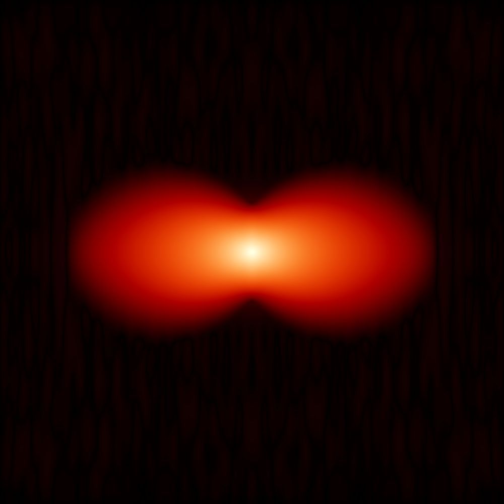
Image showing two different views of the cochlea of a human inner ear shown on a black background with bright, detailed turquoise images and violet colours. The image is very clear and detailed. It was created using the new light sheet fluorescence microscopy platform developed by researchers.
The image is adapted from Aakhte, M. et al., Nature Biotechnology, DOI: 10.1038/s41587-025-02882-8; openly licensed via CC BY 4.0
Faster, clearer, deeper 3D imaging
Researchers developed an innovative light sheet fluorescence microscopy platform, which means detailed scans of fine networks of nerves or blood vessels are possible faster: www.uni-goettingen.de/en/3240.html...
#NatureBiotechnology: doi.org/10.1038/s415...
08.01.2026 14:34 — 👍 5 🔁 3 💬 0 📌 0

Hörschnecke eines menschlichen Innenohrs sichtbar gemacht mit dem neuartigen Lichtblatt-Fluoreszenzmikroskop © Adaptiert nach Aakhte, M. et al., Nature Biotechnology, DOI: 10.1038/s41587-025-02882-8; lizensiert nach CC BY 4.0
Lichtblattmikroskope erstellen 3D-Scans von Geweben und Organen – erzeugen bei großen Proben aber unscharfe Bilder. Forschende haben eine Plattform entwickelt, die diese Hürde überwindet. Mehr erfahren: www.uni-goettingen.de/de/3240.html...
#MBexC #UMG @mbexc.bsky.social @huiskenlab.bsky.social
16.01.2026 08:34 — 👍 11 🔁 3 💬 0 📌 0

Rendering of a full cochlea (left) with three stains (PV, VGlut3, CTBP2) shown in three different colors (red, blue, cyan). The whole cochlea is a spiral shaped structure, with spiral ganglion neurons (SGNs) in the inner helix, labeled by PV and inner hair cells (IHCs) in the outer helix, labeled by Vglur3. The figure also shows zoom ins. The right hand side shows segmentation results for SGNs, IHCs (represented by colored masks) and synapse detections (represented by colored dots).
Preprint alert! CochleaNet, our framework for analyzing light-sheet data of the cochlea. It consists of three networks to segment spiral ganglion neurons, inner hair cells, and to detect synapses. See rendering of a full cochlea in the image, find the preprint at doi.org/10.1101/2025....
18.11.2025 07:58 — 👍 26 🔁 4 💬 1 📌 1
Studying cochlear neuroanatomy demands imaging the entire cochlea at subcellular resolution. In our recent study (tinyurl.com/isotropicLSFM), we recorded the cochlea at isotropic resolution across the whole organ, laying the groundwork for what follows…
#lightsheet
#isotropic
#cochlea
20.11.2025 21:20 — 👍 19 🔁 5 💬 1 📌 0
For technical details see the following post:
16.11.2025 14:47 — 👍 1 🔁 1 💬 0 📌 0
Very happy to share our latest work extending iterative immunofluorescence to in toto imaging of early zebrafish embryos (3D-4i), integrated with a 3D-dedicated image analysis pipeline! 🐟🔬📊
Huge kudos to Max Hess for this PhD milestone! 💪
@lucaspelkmans.bsky.social shorturl.at/cO0u7
Let's zoom in!
29.10.2025 14:20 — 👍 38 🔁 13 💬 4 📌 0
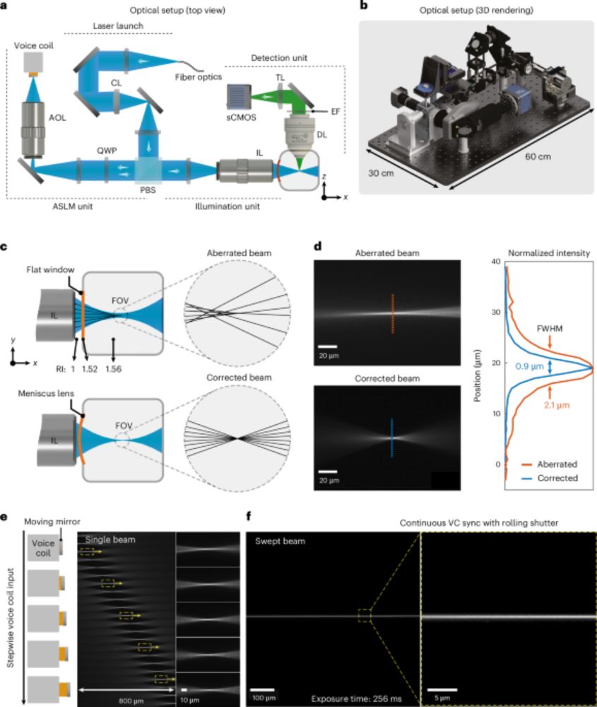
Happy to see our paper “Isotropic, aberration-corrected light sheet microscopy for rapid high-resolution imaging of cleared tissue” in @natbiotech.nature.com – with Tobias Moser’s lab from @unimedizin-goe.bsky.social and @janwenzel.bsky.social
www.nature.com/articles/s41...
#microscopy #lightsheet
14.11.2025 16:41 — 👍 18 🔁 9 💬 2 📌 1

Our image of a sea squirt oozoid (Thalia democratica) was selected as an Image of Distinction in Nikon Small World. Imaged with the Flamingo microscope by @metteh-thorsager.bsky.social at @biodev-vlfr.bsky.social in Villefranche-sur-Mer. Salp sample courtesy of Alexandre Alie.
nikonsmallworld.com/
15.10.2025 19:41 — 👍 16 🔁 4 💬 2 📌 1
The future of High-Content Imaging has arrived.
-> Ultra-fast 3D imaging of multi-well plates at high resolution using an air objective!
See previous posts and the new miOPM preprint for details: doi.org/10.1101/2025...
13.10.2025 00:44 — 👍 39 🔁 9 💬 2 📌 1
Postdoc opportunity in the Averof lab: a crustacean meets a flamingo—not on the menu, but in the light sheet. Track the origins of cells during leg regeneration in Parhyale hawaiensis using light sheet microscopy. Excited to be part of this interdisciplinary project. Details on how to apply below.
03.10.2025 15:32 — 👍 4 🔁 2 💬 0 📌 0
We present a simple method to easily increase the imageable depth of an expansion microscopy gel on a typical inverted microscope ten-fold, using some carefully placed FEP film and a water dipping objective lens:
15.09.2025 08:42 — 👍 76 🔁 32 💬 3 📌 2
The Huisken lab is now on Bluesky @huiskenlab.bsky.social - follow us for updates on our activities!
15.09.2025 15:34 — 👍 5 🔁 1 💬 0 📌 0

We brought our Flamingo microscope to the light sheet microscope course in Dresden (#LISH25). We captured extensive time-lapse recordings and are excited to share highlights from our experiments with Hydra and Zebrafish.
15.09.2025 15:26 — 👍 17 🔁 3 💬 1 📌 0
We are excited to share our development of a new 10X lens with NA=0.50 over a 7.2 mm FOV and a 35 mm working distance (water), fully corrected from 400-850 nm. This lens will power our upcoming ExA-SPIM "2" and is available for dissemination - please get in touch if interested!
19.05.2025 13:10 — 👍 46 🔁 8 💬 1 📌 2
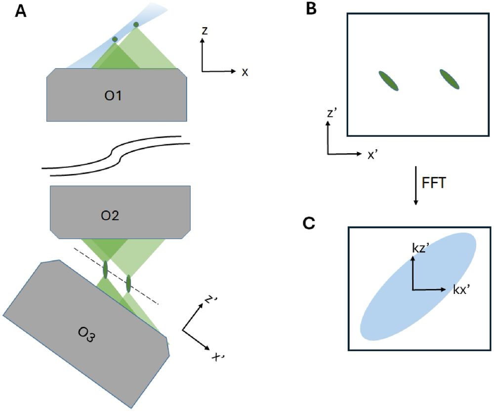
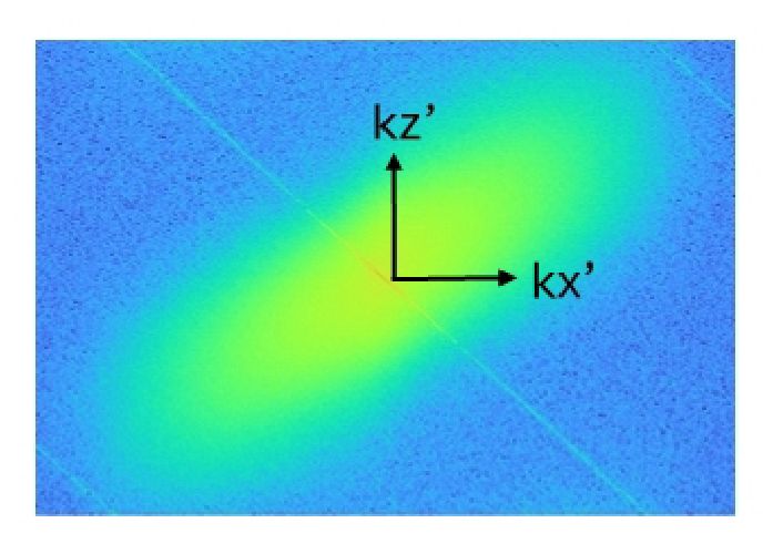
Our manuscript on accelerating the acquisition rate in oblique plane microscopy (OPM) is online now:
opg.optica.org/boe/fulltext...
Our work addresses the issue that an OPM PSF is tilted, and as such it leaves a lot of "empty space" in Fourier space (FFT of experimental data on the right)... 1/n
02.04.2025 18:21 — 👍 40 🔁 11 💬 1 📌 0
Microscope objectives can be confusing, and detailed specs (or even basic properties) are often hard to find. Here's an introductory resource to help new builders and instrument designers with the basics. It's a work in progress so feedback is appreciated:
amsikking.github.io/microscope_o...
25.02.2025 17:06 — 👍 37 🔁 14 💬 1 📌 4
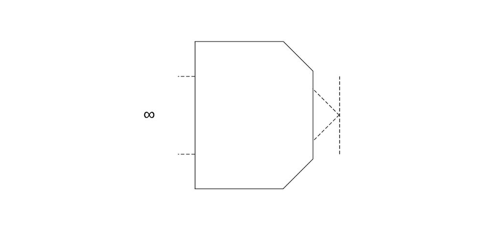
To solve the collision problem in OPM (previous post), lower numerical aperture (NA) objectives with longer working distances were used:
-> Here we show the pupils in a remote refocus (3 circles). The reduced NA in the downstream objectives loses light (yellow circle) and lowers the resolution!
22.02.2025 15:53 — 👍 1 🔁 1 💬 0 📌 0
Thanks for the kind words, Alfred!
22.02.2025 19:04 — 👍 2 🔁 0 💬 0 📌 0
Remote refocus (RR) strikes again! ;)
Congrats to @aakhte.bsky.social and team on their interesting blend of opto-mechanical innovations!
22.02.2025 18:59 — 👍 7 🔁 1 💬 1 📌 0
Researcher at the University of Göttingen, Germany. Microscopist, Method developer: super-resolution imaging, SMLM, DNA-PAINT, FLIM, Multiplexing, Microfluidics.
🇲🇽 PhD Student @MPIforBI / UniGoe interested in ethology, neural computations and evolution. NSB-MBL '25
A career network featuring science jobs in academia and industry.
Visit our platform at www.science.hr
A platform for life sciences. Publications, research protocols, news, events, jobs and more. Sign up at https://www.lifescience.net.
Science Events helps you organise a scientific conference or workshop. Use our platform to organise your next event! Visit our platform at https://www.sci.events
SNSF Ambizione junior group leader @UZH Fascinated by how embryos self-organise their shapes and patterns 🐠
Postdoc @ Pelkmans lab | PhD @ Heisenberg lab
Assistant Professor of Chemistry and Biological Sciences @UIC
Studying Neurobiology, Chemistry, Microscopy, and Nanoelectronics
Lab website: http://www.gaogroup.site/
Canada Research Chair. Misfolding proteins, chaperones and cholinergic function in neurodegeneration. Interested in reproducibility, translational research and open science. ORCID: 0000-0002-3028-5778
I also post pics of sunsets and bikes.
Evolutionary Optimization of Neuronal Processing
Comparative developmental biology, regeneration, non-conventional model organisms, live imaging; see www.averof-lab.org
Applied physicist & microscopist/microscopologist
High-end modular and portable #lightsheet microscopy for inside and outside the lab / Flamingo project / Imaging development, physiology and anatomy in live specimens and cleared tissues. Former @morgridgeinstitute.bsky.social, @mpi-cbg.de
huiskenlab.com
Publishing the best of biotech science and business. Find us on Twitter, Facebook & Instagram. Part of @natureportfolio.nature.com.
PI at HIT. We develop imaging and image-analysis solutions for biomedical applications. #sparsedeconvolution #SACDimaging #rFRCmetric #SN2N
Ph.D. | Ex-Neuro & Ephys | Microscopes & Lasers | Coherent Corp. | Opinions mine
Developing integrated technological platforms to study the molecular mechanisms that drive processes in development and disease.
@ Fiolka Lab, UT Southwestern
The official account for the Life Science Solutions division of the Nikon Healthcare Business, with a focus on biological microscopy products and services for the life science, biotech/pharma, and clinical laboratory markets.
NeuroAI, Deep Learning for neuroscience, visual system in mice and monkeys, computational lab based in Göttingen (Germany), https://sinzlab.org