Geng, @kjverhey1.bsky.social et al find that KIF1C forms condensates where the exposed #kinesin motor domains entangle nearby #microtubules, causing them to bend & break in a process dependent on motor processivity, cluster properties, cytoplasmic viscosity, & MT anchors rupress.org/jcb/article/...
11.08.2025 17:15 — 👍 49 🔁 12 💬 2 📌 3
What would happen if cells would express more molecular motors ?
They would break all microtubules 😱
As shown by recent great work from @kjverhey1.bsky.social
rupress.org/jcb/article-...
07.10.2025 15:08 — 👍 51 🔁 13 💬 0 📌 1

We present multi-immersion Oblique Plane microscope (miOPM), a light-sheet platform that can be adapted to a wide range of applications, from sensitive live cell imaging to imaging organs and cleared tissues.
www.biorxiv.org/content/10.1...
06.10.2025 17:52 — 👍 111 🔁 42 💬 5 📌 2

Playing around with @kwolbachia.bsky.social 's LUT manager plugin. Absolutely love the campfire LUT🔥 😍#microtubulemonday
06.10.2025 09:30 — 👍 21 🔁 4 💬 2 📌 0
This is incredibly beautiful, and the fact that the resolution is sufficient to follow the path of DNA spooling inside the virion head and through the tail is amazing
06.10.2025 11:41 — 👍 33 🔁 9 💬 1 📌 0
Thank you Anna!
05.10.2025 15:53 — 👍 0 🔁 0 💬 0 📌 0

RhoA (blue) and the actin cytoskeleton (magenta) are shown in a set of primary microglia.
For #FluorescenceFriday, RhoA (blue) and the actin cytoskeleton (magenta) are shown in a set of primary microglia 🔬 #Neuroscience #Microscopy
03.10.2025 13:07 — 👍 81 🔁 16 💬 4 📌 1
Thank you Shweta! 😄
05.10.2025 07:40 — 👍 0 🔁 0 💬 0 📌 0

Figure from our review paper showing various fluorescence microscopy images of cells and structures of the kidney
Sharing this excellent review now out from postdoc Sarah McLarnon! Lots of useful info on 3D imaging, quantification of imaging data, and how this has been applied to the kidney. And since it's #FluorescenceFriday, sharing a figure with our own imaging data!
www.sciencedirect.com/science/arti...
03.10.2025 23:29 — 👍 36 🔁 8 💬 0 📌 0

smFISH for immediate early gene Fos in a section of the mouse hippocampus
For #FluorescenceFriday - simple image, but structure of hippocampus is beautiful with additional fanfare. 🟢 is Fos - labeling cells that get activated in response to disinhibition of inhibitory neuron subpopulation in the prefrontal cortex. Image credit to JHU Neuroscience student Aaron Salisbury.
03.10.2025 13:08 — 👍 45 🔁 5 💬 0 📌 0

Confocal microscopy photo showing neurons and microglia
👻 "Dark Forest Within" 🧠
What if the brain is a creepy forest? Our neurons are the 🌲, old (as old as we are!), and without mobility. Suddenly, comes the microglia, our brain's immune cells. Everything lights up when they move around.
🟢microglia🟣neurons
#FluorescenceFriday 🎃
03.10.2025 21:44 — 👍 96 🔁 18 💬 4 📌 1

I’m happy to share some plugins I’ve been developping this summer: "Channels and Contrast" and LUTs Manager!
I can’t find new bugs and ideas by now so I need your help to please test them in your machines and report bugs, feedbacks and ideas! forum.image.sc/t/looking-fo...
04.10.2025 11:50 — 👍 92 🔁 34 💬 6 📌 2
Looks awesome! I can’t wait to try it out 🤩
05.10.2025 06:26 — 👍 1 🔁 0 💬 0 📌 0
Thank you! It’s a Santa Cruz antibody (26C4), diluted 1:200, and left overnight :)
05.10.2025 06:21 — 👍 1 🔁 0 💬 0 📌 0
Thanks for sharing Leah! 😁
05.10.2025 06:18 — 👍 1 🔁 0 💬 0 📌 0
Thank you! 😄
05.10.2025 06:17 — 👍 1 🔁 0 💬 0 📌 0
RhoA (blue) and the actin cytoskeleton (magenta) are shown in a set of primary microglia.
For #FluorescenceFriday, RhoA (blue) and the actin cytoskeleton (magenta) are shown in a set of primary microglia 🔬 #Neuroscience #Microscopy
03.10.2025 13:07 — 👍 81 🔁 16 💬 4 📌 1
Mario Ledesma-Terrón, Diego Pérez-Dones, David Míguez and colleagues @cbm-csic-uam.bsky.social @ifimacuam.bsky.social present OSCAR, a framework to quantify 3D stacks with high cellular density.
journals.biologists.com/jcs/article-...
03.10.2025 10:08 — 👍 73 🔁 22 💬 1 📌 1

Nanotubes everywhere between neurons! A bit skeptical about these "dendrite-dendrite nanotubes" from a phalloidin staining in culture, but let's read on… www.science.org/doi/10.1126/...
02.10.2025 18:57 — 👍 38 🔁 8 💬 1 📌 0
Easily my new favorite image of yours!! Awesome work Maik!!
03.10.2025 11:21 — 👍 2 🔁 0 💬 0 📌 0
My entry for today’s #FluorescenceFriday: a pupal #Drosophila testis with muscles expressing
🔵 lifeact &
🔴 RFP-nls
Honored & grateful to receive an honorable mention at @healthcare.nikon.com Nikon Small World 🌍🔬✨
🔗 www.nikonsmallworld.com/galleries/20...
#NikonSmallWorld #Microscopy #ScienceArt
03.10.2025 09:06 — 👍 212 🔁 40 💬 11 📌 1
See this? This = implanting mouse embryo. Usually this happens inside its mother and is invisible to us, but we can actually watch implantation ex vivo with the hope of understanding why implantation goes awry in embryos of older women. A 🧵...
01.10.2025 18:20 — 👍 219 🔁 68 💬 11 📌 18
You're watching 12 hrs of live cell imaging compressed into a GIF.
A multi-nucleated mouse pre-adipocyte tries to undergo mitosis, triggers a checkpoint because it has too much DNA, & undergoes apoptosis.
The last stage is called "blebbing", which is wonderfully descriptive.
(🔬: NanoLive Imaging)
29.09.2025 19:42 — 👍 86 🔁 18 💬 3 📌 1
Time for a thread!🧵 How different is the molecular organization of thylakoids in “higher” plants🌱? To find out, we teamed up with @profmattjohnson.bsky.social to dive into spinach chloroplasts with #CryoET ❄️🔬. Curious? ..Read on!
#TeamTomo #PlantScience 🧪 🧶🧬 🌾
elifesciences.org/articles/105...
1/🧵
25.09.2025 18:00 — 👍 135 🔁 43 💬 3 📌 6
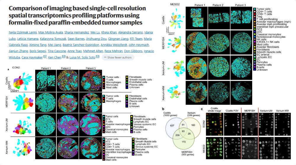
Comparing single-cell #SpatialTranscriptomics methods on FFPE human tumor #TissueMicroArray
CosMx 1k vs MERFISH 500 vs Xenium-UM/-MM 339
93 common genes
lung adenocarcinoma
pleural mesothelioma
Corr. Bulk RNAseq, GeoMx
#NatureComms 2025
www.nature.com/articles/s41...
28.09.2025 12:22 — 👍 19 🔁 5 💬 0 📌 2

A blood vessel in the brain for #FluorescenceFriday! Why am I looking at these in the microscope? (1/3)
26.09.2025 18:49 — 👍 47 🔁 14 💬 2 📌 1
Absolutely stunning image! 🤩
26.09.2025 18:16 — 👍 1 🔁 0 💬 0 📌 0

Image of a fluorescently labeled adult mouse kidney showing AQP2 staining of collecting ducts and connecting segment in green and alpha SMA staining of the arterial tree in magenta. The collecting ducts look like squiggly branches.
For this #FluorescenceFriday, a gorgeous image of an adult mouse kidney labeled with AQP2 and alpha SMA antibodies. AQP2 (green) marks the collecting duct and distal connecting segment while SMA marks the arterial tree. Courtesy of talented postdoc Sarah McLarnon.
26.09.2025 17:51 — 👍 113 🔁 32 💬 5 📌 0
#FluorescenceFriday: getting ready for #GEF25, playing with Ciarán's stunning ExM data
26.09.2025 11:48 — 👍 117 🔁 20 💬 6 📌 0
Prof at University of Michigan, Cell & Developmental Biology. Microtubules, Motor Proteins, Microscopy.
Associate Editor at Science covering plant science.
Prof of Organic Geochemistry and Sedimentology
Aspiring Immunobiologist, Undergrad at National Institute of Science Education and Research, India
🧠🔬 The Mesoscopic Integrated Neuroimaging Data (MIND) Platform revolutionizes brain research by combining Canada's most powerful MRI scanner with cutting-edge light sheet microscopy.
📍Western University, London, ON, Canada
PhD student
Brain & Neurogenesis | Evolution of the Brain | Genomics
Associate Prof. @ University of Turin | Into RNA, chromatin & stem cell adaptation in wound healing and early epithelial tumorigenesis | www.donatilab.org | Ex @cruk-ci.bsky.social @KCLstemcells
All things teammassspec/realtimechem. Survives on only the fanciest coffee and beer.
Laser capture microdissection | spatial proteomics | protein modification | atherosclerosis | inflammation and oxidation
Gut physiologist studying epithelial-immune dysfunction in the gastrointestinal tract 🧪 gutsciencelab.com
🔬 #organoids #macrophages #stemcells #ibd
Postdoc @ Kidney Genetics Group @Ong_Lab @ShefUni_ClinMed
Exploring the mechanobiology of phagocytes. Assistant prof at SFU.
CIRM Postdoc Fellow at UC Irvine: Piezo1, cell migration, neural development, high-res microscopy. Cinema lover.
PhD student at Jagiellonian University in Krakow | Interested in monocyte/macrophage biology 🧫 | monocyte/macrophage extracellular traps (METs) 🕸️| sepsis & intravital microscopy 🔬
Leveraging circadian immunity to promote healthy longevity.
Research scientist.
Science writer, mostly puttering about the garden.
she/her
ISDB is a non-profit scientific association that promotes the study of developmental biology
ASU Bioengineering
Microglial functional states
Systems neuroimmunology
Democratized biologics
Weird Synthbio
#iGEM #microglia
biophysics / cytoskeleton / evolutionary cell bio / education
Asst. Prof @ Univ. Kentucky
Past: @StearnsLab & J. Feldman Lab, @DumontLab
https://alonglab.org