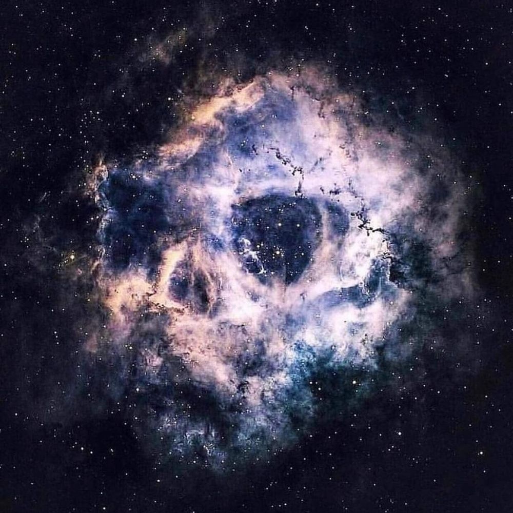
Delivery vehicles in a storm 🚚🌩️
The 🔵 is an influenza A virus storm overtaking these lung cells. The 🟠 is the AGFG1 transport vehicles braving the storm. And the 🟡 is the nucleus, struggling to keep control.
Kicking off a month-long collaboration w/ @proteintech.bsky.social
#fluorescencefriday
06.02.2026 09:10 — 👍 8 🔁 1 💬 1 📌 0
Thank you! I'm happy they are finding their audience 😁
27.01.2026 09:02 — 👍 0 🔁 0 💬 0 📌 0
Deconstructing the cell's immune system.
This is a Vero cell expressing the antiviral Human MX1 dynamin-like large GTPase (my pet protein).
It is ready to fight back, when a viral invader arrives!
🟠 MX1
🔵 Actin
⚪ DNA
#SciArt #Virology #MX1 #Microscopy #Fluorescence
27.01.2026 08:49 — 👍 4 🔁 0 💬 0 📌 0

A high-contrast fluorescence microscopy image of Vero E6 cells. The cytoskeleton (actin filaments) is stained in bright gold, appearing as a network that stretches across the cells. The nuclei are stained in cyan blue, looking like oval sapphires embedded within the gold structure. The cells appear to be gripping the surface, displaying high tension.
The Gold Standard.
These are Vero E6 cells (African Green Monkey kidney), the workhorses of virology.
They lack interferon, making them the perfect host for viral research.
Today, they are just pristine jewels.
💛 Gold: Actin
🩵 Sapphire: Nuclei
#SciArt #Virology #FluorescenceFriday
23.01.2026 10:35 — 👍 22 🔁 5 💬 1 📌 0

A fluorescence microscopy image of A549 lung cells infected with influenza A virus. The image is chaotic and colorful. The cytoplasm is filled with bright cyan viral nucleoprotein. Gold microtubules form a dense network, and pink recycling endosomes are scattered throughout. Green nuclei are visible in the center of the cells.
Project X: Cellular Edition. 🥳
This influenza infection went way too hard. 24h later, the virus (🔵) is swinging from the chandeliers (🟠 microtubules) and there are empty bottles everywhere (🩷 recycling endosomes).
Biology is beautiful chaos.
#Virology #Microscopy #Flu #SciArt #MicroscopyMonday
19.01.2026 09:34 — 👍 6 🔁 0 💬 0 📌 0
Thanks!
In these experiments there is around 20% of "virus-inside-a-virus" particles and the rest are free. This % depends on the helper virus!
And yes, free particles can have glycoproteins!
The paper has just been accepted so you'll be able to read all about it in greater detail soon! 😄
17.01.2026 09:06 — 👍 0 🔁 0 💬 2 📌 0
Imaged with a @zeiss-microscopy.bsky.social LSM989 + Airyscan 2.
16.01.2026 12:07 — 👍 0 🔁 0 💬 0 📌 0
Imaged with a @zeiss-microscopy.bsky.social LSM980.
13.01.2026 10:22 — 👍 1 🔁 0 💬 0 📌 0

High-contrast fluorescence microscopy of a mouse fibroblast cell. The cell glows in neon cyberpunk colors. Cyan actin filaments form the outer shape, magenta microtubules fill the center, and an orange DNA nucleus sits in the middle.
Biology in Cyberpunk mode.
Who said nature needs to stick to earth tones? This NIH3T3 cell has adopted the full vaporwave palette.
Turning a lab workhorse into a neon light show.
🩵 Actin
💜 Tubulin
🧡 DNA
Sometimes science just looks cool.
#SciArt #Vaporwave #Microscopy #actin #Fluorescence
13.01.2026 10:22 — 👍 14 🔁 1 💬 1 📌 0
Imaged with a @oxfordinstruments.bsky.social Dragonfly Spinning Disk Confocal microscope 🔬
08.01.2026 23:38 — 👍 1 🔁 0 💬 0 📌 0
The Big Crunch.
The Big Bang.
And the expansion that follows.
Not a simulation of the early universe, but a single A549 cell dividing.
Watch the Rab11a endosomes rush like a supply fleet. One life becoming two.
Cosmology on a cellular scale.
#SciArt #Microscopy #Fluorescencefriday #BigBang
08.01.2026 23:37 — 👍 5 🔁 0 💬 1 📌 0
🔬 Imaged with a @zeiss-microscopy.bsky.social LSM980 + Airyscan 2.
07.01.2026 08:30 — 👍 1 🔁 0 💬 0 📌 0

High-contrast fluorescence microscopy of a human fibroblast cell. A golden web of actin filaments radiates outward like a starburst, forming angles, parallels and geometric stars. Cyan septin structures form scaffolds. A grey DNA nucleus sits in the center. A snapshot of the chaotic beauty, found at the center of biology.
At first glance, chaos.
Looking closer at this human fibroblast however, angles, parallels, and geometric stars appear.
Order in biology is often disguised as chaos. Beautiful chaos.
A golden web spun from the fabric of life.
💛 Gold: Actin
🩵 Cyan: Septin 7
🩶 Grey: DNA
#SciArt #Microscopy
07.01.2026 08:30 — 👍 15 🔁 3 💬 1 📌 0

Boa Constrictor Brain Cells Slither into Focus
From reptiles and Tasmanian devils to humans, one researcher zooms in on unseen features of the brain and liver cells, like the cytoskeleton.
@viroscope.bsky.social (@karimaj.bsky.social team) partage dans The Scientist ses travaux sur les deltavirus et l’imagerie de cellules, des cellules humaines aux cellules de boa constrictor 🐍.
👉 Lire l’article : “Boa Constrictor Brain Cells Slither into Focus”
www.the-scientist.com/boa-constric...
13.10.2025 11:13 — 👍 9 🔁 7 💬 0 📌 0
Thanks so much! That means so much to me 🤩
Working with MX1 proteins was definitely not easy, but coming across this mechanism felt super rewarding!
My question now is: what about the mechanisms behind the early stage blocks of the avian flus? 😄
08.10.2025 12:24 — 👍 2 🔁 0 💬 0 📌 0
Go and vote for @dianabrychka.bsky.social's stunning image of a brain organoid ! 🤩
30.06.2025 13:14 — 👍 5 🔁 1 💬 0 📌 0
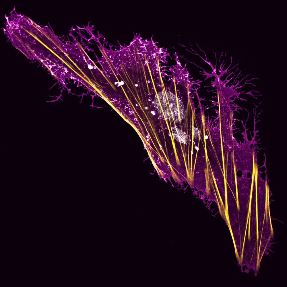
A Boa Constrictor brain-derived cell expressing a viral glycoprotein (magenta) and stained for actin (yellow) and DNA (white).
#FluorescenceFriday
27.06.2025 11:20 — 👍 79 🔁 16 💬 1 📌 0
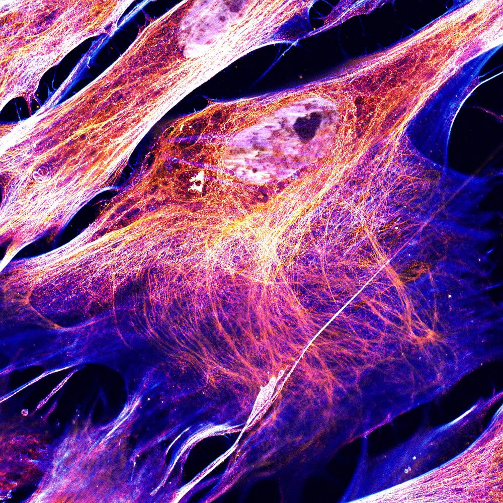
HFF cells stained for DNA ⚪, intermediate filaments 🟠, microtubules 🔴 and actin 🔵.
#FluorescenceFriday
#SciArt
30.05.2025 14:18 — 👍 29 🔁 6 💬 1 📌 0
Aha thank you for the compliment!
23.05.2025 15:43 — 👍 0 🔁 0 💬 0 📌 0
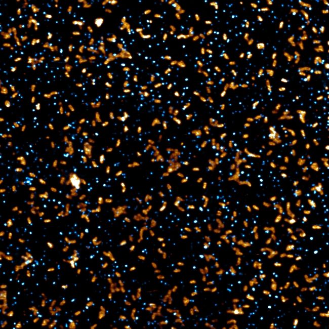
For #FluorescenceFriday, a field of individual VSV (rod-shaped, in 🟧) and Deltavirus (dot-shaped, in 🔵) particles imaged by STED (20nm per pixel).
We now know that Deltaviruses can package inside particles of other viruses.
Can you find any of these dual particles in this field?
23.05.2025 12:05 — 👍 20 🔁 2 💬 2 📌 0
Beautiful image!
16.05.2025 17:40 — 👍 3 🔁 0 💬 1 📌 0

🔬✨ Today for #FluorescenceFriday:
A close-up of the dorsal part of the first gill arch in a mature zebrafish.
The intricate vascular network supporting respiration, visualized using kdrl:mcherry.
#Zebrafish #DevBio #Microscopy #Fluorescence #SciArt
16.05.2025 13:38 — 👍 50 🔁 11 💬 4 📌 2

Viruses in the forest of the lungs.
Human airway epithelia stained for actin 🟢 and SARS-CoV-2 RNA 🟣.
#FluorescenceFriday
#SciArt
16.05.2025 11:01 — 👍 15 🔁 0 💬 0 📌 0
Thank you for sharing our work! It was great discussing with you at #ECV2025 !
12.05.2025 13:39 — 👍 1 🔁 0 💬 0 📌 0
Saw this presented at ECV 2025. Fanastic and thought provoking work! Congrats to @viroscope.bsky.social, @karimaj.bsky.social and coauthors.
10.05.2025 10:45 — 👍 8 🔁 4 💬 1 📌 0
Deltaviruses spread through a viral Trojan Horse https://www.biorxiv.org/content/10.1101/2025.05.09.653040v1
10.05.2025 03:17 — 👍 2 🔁 1 💬 0 📌 0
Deltaviruses spread through a viral Trojan Horse https://www.biorxiv.org/content/10.1101/2025.05.09.653040v1
10.05.2025 03:17 — 👍 2 🔁 1 💬 0 📌 0
📌🇨🇭 / 🗣️🇫🇷 🇬🇧 🇩🇪
⚠️Skeptical inside🧐Rational Rebel
👨🔬 #PhD in the making (neuro)
❤️#sciences , toute curiosité, gros plus pour l'imagerie🔭 📸 🔬 🖼️ 🗺️!
🎮 : KSA/P🚀 GW2 🧙♂️ MLBB 🌈
Géo-localisations sur: @geoloc-holmes.bsky.social
#whereisthis #Thenandnow
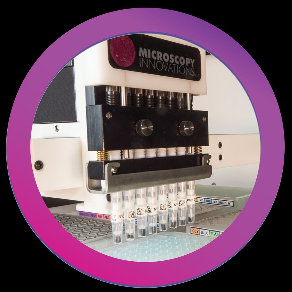
Microscopy Innovations provides benchtop lab automation to simplify and streamline sample prep for electron microscopy. Its mPrep™ASP automated specimen processor is widely acclaimed in leading microscopy laboratories.
https://giacobarto.github.io/
I love biophysics, jazz, poetry, and activism, especially when mixed together!
Into phase separation, and active nematics.
JdC postdoc at University of Barcelona, previously at MPI PKS Dresden and Uni Augsburg
Postdoc at Dey|Dudin|Schwab Groups, EMBL
Mitosis and MTOCs in Ichthyosporea, fungi and more
PostDoc at the i3S in Porto. Interested in the spatial and temporal dynamics of cell division. Sometimes I draw stuff.
Cell biologist 🔬 | Exploring the mysteries of centrosomes and cytoskeleton diversity🐉🦠 | Postdoc at Centriole Lab, University of Geneva
AFM expert at Nanosurf.
Follow #fridayAFM for fun experiments.
PhD in Biology of Interactions | Microbiology | Microscopy | Statistics
Associate Editor at The Scientist.
Microbe enthusiast & plant lover.
Stories: https://www.the-scientist.com/author/laura-tran-phd
Laboratoire plurithématique en biologie et santé du @cnrs.bsky.social
et de Université de Montpellier
Plurithematic laboratory in biology and health from @CNRS
and @umontpellier
www.igmm.cnrs.fr
Faculty at ICMR-NIRRCH 🇮🇳 Developmental biologist, embryo implantation, placentation and sex determination
We also develop organ on chip and organoids models of ♀️ rep tract and placenta
#womenshealth #gender #hoxgenes
🏳️🌈
Executive Editor/Team Lead Open Access Science & Medicine Journals Sage Publishing
Opinions = mine
http://linkedin.com/in/jlovick-editor
#oncology #cancerresearch #medicine #biology #cardiology #neurology #microbiology #publichealth #healthcare
Research assistant professor PI, Cross Flavi Lab @Pitt | Flavivirus antibody response & cross-flavivirus interactions | Epidemiology & Capacity Building | UC Berkeley affiliate
We make cool tabletop items :D
MSCA Postdoc fellow, Van Breusegem Lab, Oxidative Stress Signalling. VIB-UGent Center for Plant Systems Biology