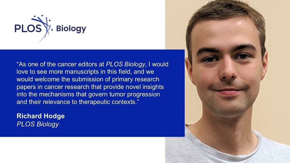
The image features a message from Richard Hodge, a cancer editor at PLOS Biology, inviting submissions of primary research papers that offer novel insights into tumor progression and therapeutic contexts, displayed alongside the PLOS Biology logo and a photograph of Richard Hodge.
In this new #Biologue post, we speak to Richard Hodge, a cancer editor at #PLOSBiology about the new Collection “Unveiling cancer crosstalk across spatial and temporal scales”, his background in #CancerResearch and his vision for #cancer biology at the journal 🧪
plos.io/3U6GXFW
01.08.2025 16:20 — 👍 1 🔁 0 💬 0 📌 0

The image features a quote from Yibin Kang of Princeton University emphasizing the need for an integrated approach to understanding cancer, displayed alongside the PLOS Biology logo and a photograph of Yibin Kang.
Cancer evolves through dynamic exchanges with its environment, harnessing these interactions to grow and adapt. @yibinkang.bsky.social introduces a new collection of articles that explore crosstalk between #cancer and its environment across temporal and spatial scales 🧪
plos.io/4moogJS
01.08.2025 16:19 — 👍 1 🔁 1 💬 0 📌 0
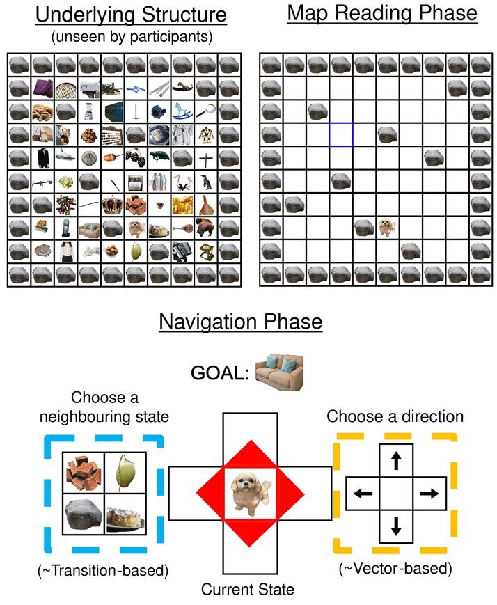
Task design and experimental set-up. Top left: underlying structure of the 8 × 8 grid, unseen by participants. Every state is represented by an image of an object, and these objects and their positions change on every trial. Top right: schematic diagram of the ‘map reading’ phase of each trial. Participants see a top–down view of the grid with objects obscured and successively click on blue squares to reveal ‘landmark’ objects at the location. After 16 clicks have been completed, a yellow square appears. Clicking on the yellow square reveals the ‘goal’ object for the trial. Bottom: schematic diagram of the navigation phase of each trial. Participants start in a random, previously unobserved location and are tasked with navigating to the ‘goal’ object they had just learnt about (displayed at the top). They can navigate in two ways. First, they could choose a direction to travel in by clicking on the corresponding arrow (highlighted yellow). This is analogous to using a ‘vector-based’ strategy. Alternatively, they could choose an adjacent state to travel to by clicking on one of the associated images (displayed in a random order; highlighted blue). This corresponds to using a ‘transition-based’ navigation strategy.
How do humans navigate unfamiliar environments? @denislan.bsky.social @lhuntneuro.bsky.social @summerfieldlab.bsky.social show that humans & deep meta-learning networks combine ‘vector-based’ & ‘transition-based’ strategies for flexible navigation in similar ways @plosbiology.org 🧪 plos.io/45uSwNm
01.08.2025 14:58 — 👍 2 🔁 0 💬 0 📌 0
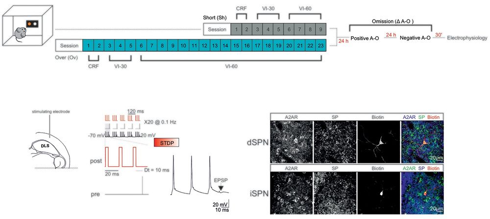
Top: Schematic of the behavioral regimes followed by ex vivo electrophysiology. Bottom left: Experimental configuration in horizontal brain slices containing the DLS. Bottom middle: The post-pre pairing protocol (negative STDP) for the induction of t-LTD. Bottom right: Confocal laser scanning microscopy images of triple immunofluorescence for adenosine A2A receptor (A2AR), substance P (SP), and biotin in patch-recorded neurons (scale bar: 20 µm).
Instrumental actions rely on flexible, goal-directed #behavior, but can become more rigid & habit-like with repetition. @vincentpb.bsky.social &co show that decreased adaptive signaling capacity of #striatal mGluR1/5 is key to this loss of #BehavioralFlexibility @plosbiology.org 🧪 plos.io/3JaRLAk
01.08.2025 14:58 — 👍 1 🔁 1 💬 0 📌 0
The image features a message from Richard Hodge, a cancer editor at PLOS Biology, inviting submissions of primary research papers that offer novel insights into tumor progression and therapeutic contexts, displayed alongside the PLOS Biology logo and a photograph of Richard Hodge.
In this new #Biologue post, we speak to Richard Hodge, a cancer editor at #PLOSBiology about the new Collection “Unveiling cancer crosstalk across spatial and temporal scales”, his background in #CancerResearch and his vision for #cancer biology at the journal 🧪
plos.io/3U6GXFW
01.08.2025 12:46 — 👍 1 🔁 0 💬 0 📌 0
The image features a quote from Yibin Kang of Princeton University emphasizing the need for an integrated approach to understanding cancer, displayed alongside the PLOS Biology logo and a photograph of Yibin Kang.
Cancer evolves through dynamic exchanges with its environment, harnessing these interactions to grow and adapt. @yibinkang.bsky.social introduces a new collection of articles that explore crosstalk between #cancer and its environment across temporal and spatial scales 🧪
plos.io/4moogJS
01.08.2025 12:45 — 👍 1 🔁 0 💬 0 📌 0
A new #cancer collection @plosbiology.org explores cancer crosstalk across temporal and spatial scales, & the power of emerging #technologies to drive our understanding and treatment of the disease.
Thanks @yibinkang.bsky.social for your strategic vision and insight!
🧪 plos.io/4moogJS
01.08.2025 09:23 — 👍 3 🔁 1 💬 0 📌 0
The image features a message from Richard Hodge, a cancer editor at PLOS Biology, inviting submissions of primary research papers that offer novel insights into tumor progression and therapeutic contexts, displayed alongside the PLOS Biology logo and a photograph of Richard Hodge.
In this new #Biologue post, we speak to Richard Hodge, a cancer editor at #PLOSBiology about the new Collection “Unveiling cancer crosstalk across spatial and temporal scales”, his background in #CancerResearch and his vision for #cancer biology at the journal 🧪
plos.io/3U6GXFW
01.08.2025 08:38 — 👍 3 🔁 3 💬 0 📌 1
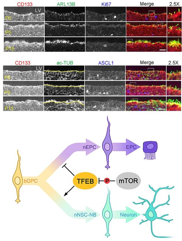
Top: CD133 showed specific enrichment on the apical surface of radial glial cells (RGCs) in the developing ventricular zone (VZ) from postnatal day 0 (P0) to P15. Ciliary membrane marker (ARL13B) and the ciliary microtubule marker acetylated-tubulin (ac-TUB) indicated ependymal cell (EPC)-lineage differentiation. Cell proliferating marker (Ki67) and neural stem cell (NSC)-lineage marker (ASCL1) indicated NSC-lineage transition. The framed region was further magnified to show details. The boundary between lateral ventricle (LV) and VZ was denoted by the yellow dotted line for better distinguishing ciliary ac-TUB and cytoplasmic ac-TUB in (B). The scale bars are 20 μm. Bottom: A model illustrating how TFEB functions in the process of bGPC specification during VZ development.
#NeuralStemCells (NSCs) & ependymal cells (ECs) are derived from #RadialGlialCells. This study uses #scRNAseq to characterize cell fate trajectories in the developing #VentricularZone, identifying TFEB as a regulator of the NSC/EPC balance @plosbiology.org 🧪 plos.io/3HbrsJG
01.08.2025 08:29 — 👍 3 🔁 0 💬 0 📌 0
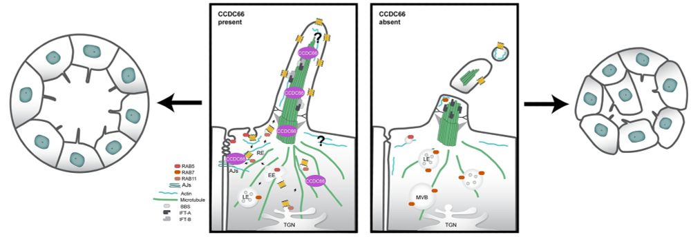
Model for cellular functions and mechanisms of CCDC66. CCDC66 is a microtubule-stabilizing protein that maintains cilium integrity and size and facilitates vesicular transport at the ciliary base. Depletion of CCDC66 results in variations in cilia size due to compromised axonemal integrity, intraflagellar transport, and increased ectocytosis events involving the release of vesicles and fragments. CCDC66 may also support ciliary ectocytosis and length stability by regulating the actin cytoskeleton. Furthermore, disruptions in both microtubule stability and the actin cytoskeleton impair RAB11 and RAB5-mediated endocytosis and membrane recycling, affecting cell adhesion and the targeting of ciliary receptors. This disruption subsequently activates protein degradation pathways through RAB7-regulated late endocytosis, potentially involving RAB7 translocation to cilia to support ectocytosis. Collectively, these defects in protein transport, cilia function, and cell contacts contribute to compromised epithelial tissue integrity and multilumen formation in kidney tubules.
The #PrimaryCilium dynamically assembles & disassembles, but how is this controlled? @cytolabkoc.bsky.social shows that #ciliopathy linked protein Ccdc66 regulates both #cilium stability & length in epithelial cells via microtubules, actin & vesicular trafficking @plosbiology.org 🧪 plos.io/4mdTS4A
01.08.2025 08:28 — 👍 6 🔁 1 💬 0 📌 0
Task design and experimental set-up. Top left: underlying structure of the 8 × 8 grid, unseen by participants. Every state is represented by an image of an object, and these objects and their positions change on every trial. Top right: schematic diagram of the ‘map reading’ phase of each trial. Participants see a top–down view of the grid with objects obscured and successively click on blue squares to reveal ‘landmark’ objects at the location. After 16 clicks have been completed, a yellow square appears. Clicking on the yellow square reveals the ‘goal’ object for the trial. Bottom: schematic diagram of the navigation phase of each trial. Participants start in a random, previously unobserved location and are tasked with navigating to the ‘goal’ object they had just learnt about (displayed at the top). They can navigate in two ways. First, they could choose a direction to travel in by clicking on the corresponding arrow (highlighted yellow). This is analogous to using a ‘vector-based’ strategy. Alternatively, they could choose an adjacent state to travel to by clicking on one of the associated images (displayed in a random order; highlighted blue). This corresponds to using a ‘transition-based’ navigation strategy.
How do humans navigate unfamiliar environments? @denislan.bsky.social @lhuntneuro.bsky.social @summerfieldlab.bsky.social show that humans & deep meta-learning networks combine ‘vector-based’ & ‘transition-based’ strategies for flexible navigation in similar ways @plosbiology.org 🧪 plos.io/45uSwNm
01.08.2025 08:27 — 👍 8 🔁 2 💬 0 📌 1
Top: Schematic of the behavioral regimes followed by ex vivo electrophysiology. Bottom left: Experimental configuration in horizontal brain slices containing the DLS. Bottom middle: The post-pre pairing protocol (negative STDP) for the induction of t-LTD. Bottom right: Confocal laser scanning microscopy images of triple immunofluorescence for adenosine A2A receptor (A2AR), substance P (SP), and biotin in patch-recorded neurons (scale bar: 20 µm).
Instrumental actions rely on flexible, goal-directed #behavior, but can become more rigid & habit-like with repetition. @vincentpb.bsky.social &co show that decreased adaptive signaling capacity of #striatal mGluR1/5 is key to this loss of #BehavioralFlexibility @plosbiology.org 🧪 plos.io/3JaRLAk
01.08.2025 08:26 — 👍 2 🔁 1 💬 0 📌 0
The image features a quote from Yibin Kang of Princeton University emphasizing the need for an integrated approach to understanding cancer, displayed alongside the PLOS Biology logo and a photograph of Yibin Kang.
Cancer evolves through dynamic exchanges with its environment, harnessing these interactions to grow and adapt. @yibinkang.bsky.social introduces our new collection of articles that explore crosstalk between #cancer and its environment across temporal and spatial scales 🧪
plos.io/4moogJS
01.08.2025 08:25 — 👍 3 🔁 2 💬 0 📌 1
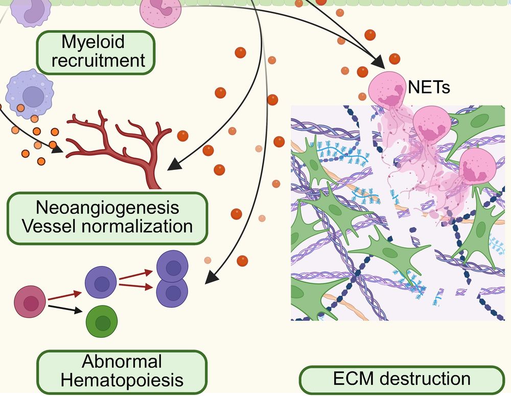
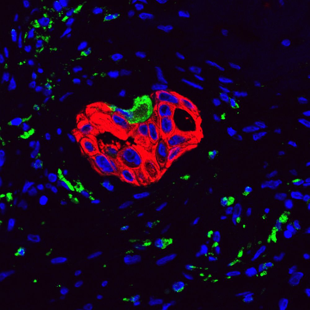
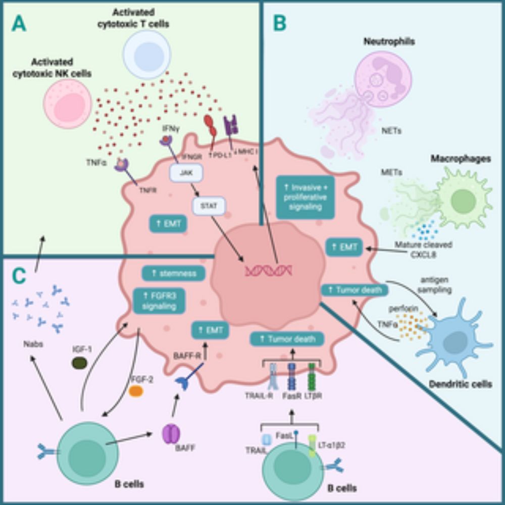
The image illustrates biological processes including myeloid recruitment, neoangiogenesis and vessel normalization, abnormal hematopoiesis, and extracellular matrix (ECM) destruction involving neutrophil extracellular traps (NETs).
In this #Essay Xiang Zhang & Fengshuo Liu explore the use of single-cell technologies to understand #cancer metastatic niches, and investigate how to harness insights gained to inform novel therapies 6/9
plos.io/4lg2yXC
01.08.2025 08:05 — 👍 1 🔁 0 💬 1 📌 0

The image depicts two rows of tall glass cylinders containing DNA double helix structures, arranged to form a symmetrical, reflective corridor that extends into the distance. Image by PublicDomainPictures from Pixabay
In this #Perspective Zhi-Ming Shao &co look at how #MultiOmics data be leveraged to help patients with #cancer in the clinic as technology continues to push forwards 3/9
plos.io/3Ja51oQ
01.08.2025 08:05 — 👍 1 🔁 0 💬 1 📌 0
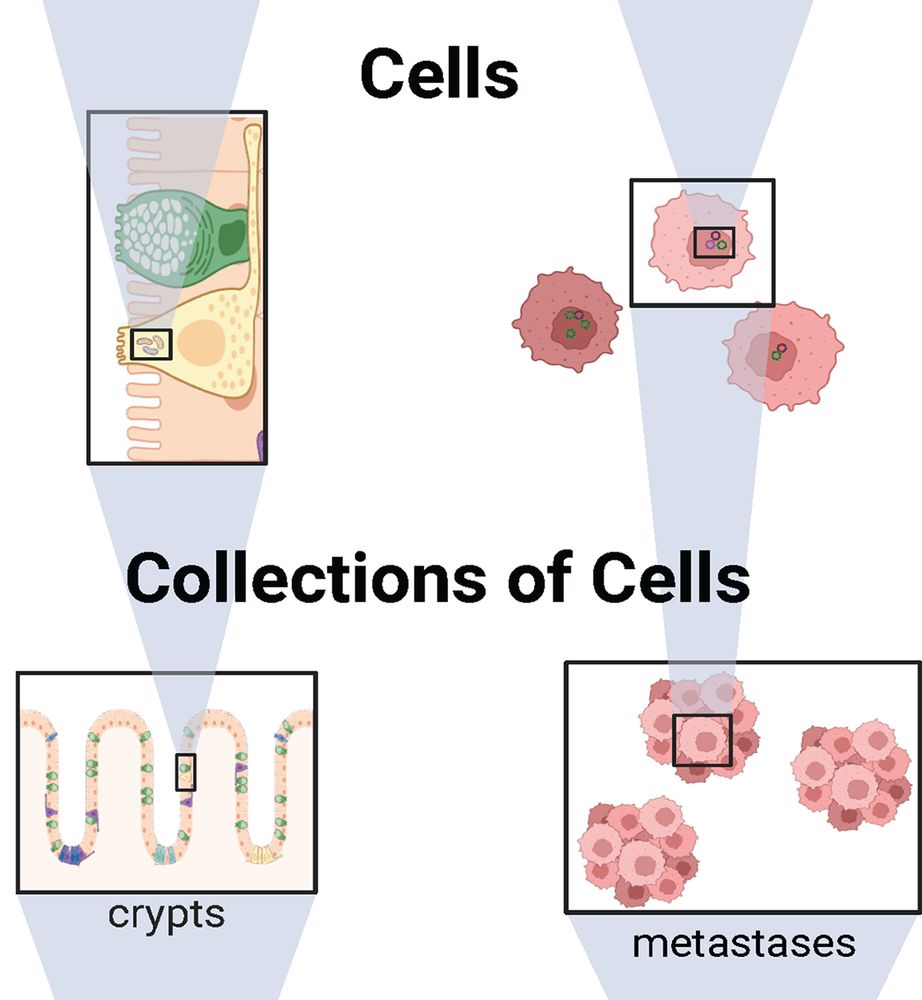
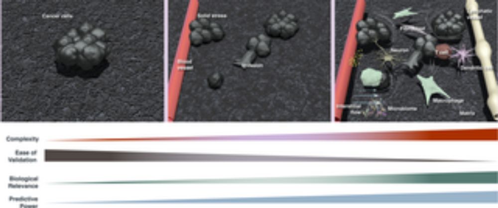
The image presents a comparison between individual cells and groups of cells, focusing on structures such as crypts and metastases.
In this #Essay, @cmaley.bsky.social @jeffreytownsend.bsky.social @lucielaplane.bsky.social @plutynski.bsky.social &co examine how evolutionary selection operates across levels, highlighting how this understanding can be used to improve #cancer prevention and treatment 2/9
plos.io/47epeUq
01.08.2025 08:05 — 👍 2 🔁 1 💬 1 📌 0
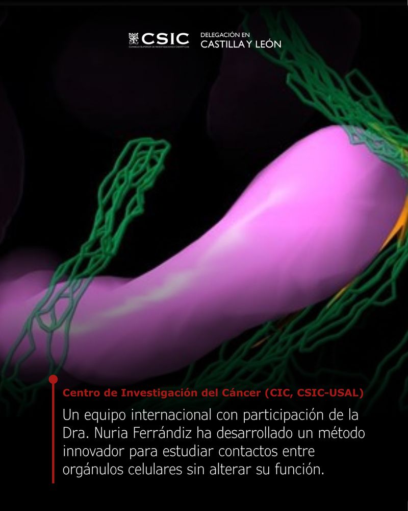
➕ La revista @plosbiology.org publica el artículo de un consorcio internacional de científicos, entre ellos @nuriaferrandiz.bsky.social (@ciccancer.bsky.social), que realiza un importante avance para la biología celular 👉 www.cicancer.org/press-room/c...
31.07.2025 18:09 — 👍 3 🔁 4 💬 1 📌 0
Task design and experimental set-up. Top left: underlying structure of the 8 × 8 grid, unseen by participants. Every state is represented by an image of an object, and these objects and their positions change on every trial. Top right: schematic diagram of the ‘map reading’ phase of each trial. Participants see a top–down view of the grid with objects obscured and successively click on blue squares to reveal ‘landmark’ objects at the location. After 16 clicks have been completed, a yellow square appears. Clicking on the yellow square reveals the ‘goal’ object for the trial. Bottom: schematic diagram of the navigation phase of each trial. Participants start in a random, previously unobserved location and are tasked with navigating to the ‘goal’ object they had just learnt about (displayed at the top). They can navigate in two ways. First, they could choose a direction to travel in by clicking on the corresponding arrow (highlighted yellow). This is analogous to using a ‘vector-based’ strategy. Alternatively, they could choose an adjacent state to travel to by clicking on one of the associated images (displayed in a random order; highlighted blue). This corresponds to using a ‘transition-based’ navigation strategy.
How do humans navigate unfamiliar environments? @denislan.bsky.social @summerfieldlab.bsky.social &co show that humans & deep meta-learning networks combine ‘vector-based’ & ‘transition-based’ strategies for flexible navigation in similar ways @plosbiology.org 🧪 plos.io/45uSwNm
31.07.2025 17:28 — 👍 3 🔁 3 💬 0 📌 0
Top: CD133 showed specific enrichment on the apical surface of radial glial cells (RGCs) in the developing ventricular zone (VZ) from postnatal day 0 (P0) to P15. Ciliary membrane marker (ARL13B) and the ciliary microtubule marker acetylated-tubulin (ac-TUB) indicated ependymal cell (EPC)-lineage differentiation. Cell proliferating marker (Ki67) and neural stem cell (NSC)-lineage marker (ASCL1) indicated NSC-lineage transition. The framed region was further magnified to show details. The boundary between lateral ventricle (LV) and VZ was denoted by the yellow dotted line for better distinguishing ciliary ac-TUB and cytoplasmic ac-TUB in (B). The scale bars are 20 μm. Bottom: A model illustrating how TFEB functions in the process of bGPC specification during VZ development.
#NeuralStemCells (NSCs) & ependymal cells (ECs) are derived from #RadialGlialCells. This study uses #scRNAseq to characterize cell fate trajectories in the developing #VentricularZone, identifying TFEB as a regulator of the NSC/EPC balance @plosbiology.org 🧪 plos.io/3HbrsJG
31.07.2025 17:10 — 👍 4 🔁 1 💬 0 📌 0
Model for cellular functions and mechanisms of CCDC66. CCDC66 is a microtubule-stabilizing protein that maintains cilium integrity and size and facilitates vesicular transport at the ciliary base. Depletion of CCDC66 results in variations in cilia size due to compromised axonemal integrity, intraflagellar transport, and increased ectocytosis events involving the release of vesicles and fragments. CCDC66 may also support ciliary ectocytosis and length stability by regulating the actin cytoskeleton. Furthermore, disruptions in both microtubule stability and the actin cytoskeleton impair RAB11 and RAB5-mediated endocytosis and membrane recycling, affecting cell adhesion and the targeting of ciliary receptors. This disruption subsequently activates protein degradation pathways through RAB7-regulated late endocytosis, potentially involving RAB7 translocation to cilia to support ectocytosis. Collectively, these defects in protein transport, cilia function, and cell contacts contribute to compromised epithelial tissue integrity and multilumen formation in kidney tubules.
The #PrimaryCilium dynamically assembles & disassembles, but how is this controlled? @cytolabkoc.bsky.social shows that #ciliopathy linked protein Ccdc66 regulates both #cilium stability & length in epithelial cells via microtubules, actin & vesicular trafficking @plosbiology.org 🧪 plos.io/4mdTS4A
31.07.2025 17:10 — 👍 6 🔁 2 💬 0 📌 0
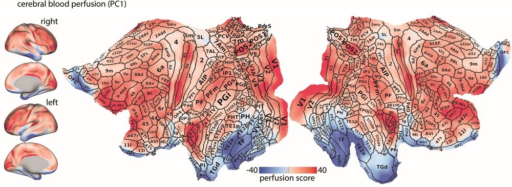
Normative blood perfusion map on lateral and medial views of the inflated and 2D flat cortical surfaces (fsLR). Borders and areal names of the multi-modal Glasser parcellation are overlaid on the flat surface. The figure highlights the non-uniform distribution of perfusion scores across the cortex, with areas that have sharp gradient compared to their underlying perfusion score levels (e.g., LIPv and MT/MST).
How does cerebral #BloodPerfusion map onto micro-, meso- & macro-scale brain structure? @asafarahani.bsky.social @misicbata.bsky.social &co characterize blood perfusion in the #HumanBrain, revealing how it changes with age & in #NeurodegenerativeDisease @plosbiology.org 🧪 plos.io/46AEURS
31.07.2025 17:09 — 👍 5 🔁 2 💬 0 📌 0
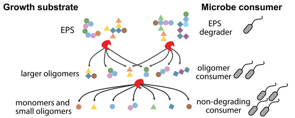
Graphical illustration of how EPS is enzymatically degraded in multiple steps into smaller fragments, fueling the growth of non-degrading species: EPS degraders break down EPS into larger oligomers for oligomer consumers; further degradation into monomers and small oligomers supports non-degrading consumers.
What is the role of extracellular polymeric substances (EPS) in carbon exchange among microbial species? @sammy-pontrelli.bsky.social &co show that #EPS, formed via #chitin degradation, drives #MicrobialDiversity by acting as a sequentially degraded #CarbonSource @plosbiology.org 🧪 plos.io/3J9kbuu
31.07.2025 17:08 — 👍 4 🔁 1 💬 0 📌 0
University lecturer, scientist, and critical thinker.
scientist studying centrosomes, cilia and centriolar satellites; associate prof @kocuniversity; alumna @ucberkeley @stanford; mom of three boys and a girl; 2X ERC StG LS3; Instagram: cellbiologist_enf
Head of Genome Biology Dept @EMBL,
Scientist, Principal investigator, Professor
Exploring genome regulation during development,
and everything to do with enhancers.
3D genome, chromatin topology,
cell_fate, embryonic Development,
Single Cell genomics
Mitochondrial Biologist, Science Enthusiast, Eternal Optimist.
Postdoc at Wu Tsai Institute, Yale University. Formerly a PhD student in neuroscience at IISc Bangalore
SMBE fosters communication among molecular evolutionists and advances the field.
🔗 smbe.org
💬 smbe2025.scimeeting.cn • @smbe-chile2025.bsky.social
👥 @smbe-idea.bsky.social
📙 @molbioevol.bsky.social • @genomebiolevol.bsky.social
Biologist studying plant-microbiota at UvA, Amsterdam. Cat person and camper.
Cognitive Neuroscientist in Amsterdam
proactivebrainlab.com
Plant / microbe biologist. PhD UNC-CH (USA), postdoc MPI for Biology (Germany), group leader SLU Uppsala (Sweden)
www.LundbergLand.org
Neuroscientist | Group leader at Marseille Developmental Biology Institute, CNRS & Aix-Marseille Université
https://www.ibdm.univ-amu.fr/team/local-translation-in-axon-regeneration/
Researcher at Cold Spring Harbor Laboratory; Interests: Neuroscience, Predictive processing, Olfaction; albeanulab.cshl.edu
Bacterial geneticist interested in host-pathogen interactions. Associate Professor at Emory University School of Medicine. Dept. of Microbiology & Immunology
Zoologist, reproductive biologist, adventure seeker and nature lover.
We’re a virology and chromatin lab digging into how Epstein-Barr Virus (EBV) rewires the host cell to cause cancer. We mix epigenetics, 3D genome biology, and metabolism to figure out how it all works—and how to stop it.
🇲🇽🇯🇵
Computer Vision and Machine learning engineer at
CZ Biohub SF || Ph.D || views are my own || (he/him)
Neuroscientist at Collège de France and @Inserm, Interest in #sleep mechanisms, #neurone, #astrocyte, #electrophysiology
#BAW #Alice2630 #FENS #glia #CdF1530 #SdC2025 #FPC2025
Plant evolutionary genomics
Retired librarian. Interested in science (mainly biomedical), choral music, running, countryside, libraries (of course), literature, the Philippines, LGBTQIA+, neurodivergence.
Science communication and anything else I happen to like.
Digital content officer at the Francis Crick Institute