Are you ready to share your science with the world? ISD Travel Awards provide financial support to help members present their work in cell and developmental biology at major scientific meetings.
Apply now to expand your reach: https://bit.ly/4gp0go3
The deadline is Feb 28.
22.01.2026 00:21 — 👍 1 🔁 0 💬 0 📌 0

Fig. 1. Conservation of zinc finger domains of Osr1 and Osr2 in vertebrates. (A) Amino acid sequence alignment of Osr1 using MafftWS sequence alignment reveals high conservation of the zinc finger binding domains. B) Amino acid sequence alignment of Osr2 using MafftWS sequence alignment reveals high conservation of zinc fingers 1–3 in all species. Zinc fingers 4 and 5 are only conserved in chick, mouse, and humans. Zinc finger domains 1–3 and 4–5 are indicated by red and purple bars, respectively, shown above the amino acid sequence of interest. OSR2B, the alternatively spliced protein product has only been identified in mammals and was therefore omitted from the protein conservation analysis. Conservation is depicted below the corresponding amino acid sequence.
“Odd-skipped family members have conserved roles in segmentation, appendage, excretory system and gut development in bilaterian animals,” by Vasikar Murugapoopathy et al. Read the open access Perspectives article in Differentiation: https://doi.org/10.1016/j.diff.2025.100930
20.01.2026 17:09 — 👍 2 🔁 0 💬 0 📌 0

Fig. 1. Illustration of the triad system in tissue engineering, representing the interaction between cell types, scaffolds, and external factors.
Review article: Siti Nurnasihah Md Hashim et al. explored how chemical and mechanical microenvironments cooperate to guide epithelial commitment across both conventional 2D and advanced 3D culture models. Learn more: https://doi.org/10.1016/j.diff.2026.100931
13.01.2026 17:14 — 👍 1 🔁 0 💬 0 📌 0

New to Differentiation: Explore the curated collection of 11 articles highlighting the latest key advancements and discoveries in osteo biology—from 2023 to present: https://www.sciencedirect.com/special-issue/10DXVD79TL0
10.12.2025 17:15 — 👍 2 🔁 0 💬 0 📌 0
Parth Aphale, Himanshu Shekhar, and Shashank Dokania provide a critical appraisal of “Osteocyte-like differentiation of osteosarcoma by inorganic phosphate,” by Suzuki et al. (Differentiation, 2025, 146:100912). Read the letter to the editor to learn more: https://doi.org/10.1016/j.diff.2025.100915
04.12.2025 21:24 — 👍 1 🔁 0 💬 0 📌 0
Deadline approaching: Submit to the forthcoming Differentiation special issue, “Splicing in Development and Disease,” by December 30, 2025! Learn more about the scope, guest editors, and benefits of publishing in a special issue: https://bit.ly/4o6TAxW
01.12.2025 16:45 — 👍 2 🔁 0 💬 0 📌 0

Fig. 2. Dermal bone mineralization was not generally accelerated in fam20b−/− embryos.
“fam20b-dependent proteoglycans do not affect dermal bone formation and fin regeneration, but Bmp signalling promotes fin regenerate outgrowth,” by Elham Koosha et al. Read the open access article in Differentiation to learn more: https://doi.org/10.1016/j.diff.2025.100914
25.11.2025 16:45 — 👍 3 🔁 0 💬 0 📌 0

Screenshot of presentation titled "Enteric neural crest development in Astyanax mexicanus surface fish and cavefish"
Thank you, everyone, who joined the latest “Biological Differentiation Across the Scales” virtual seminar last week with Pavani P. Perera from Misty Riddle’s lab at @unevadareno.bsky.social. Couldn’t attend the live seminar? The recording is now available online! Watch now: https://bit.ly/4oIeT9k 🐟
21.11.2025 19:50 — 👍 2 🔁 0 💬 0 📌 2

Biological Differentiation Across the Scales • November 14, 2025, at 12:00 p.m. Central • Register Now
TOMORROW: Join the Editors-in-Chief of Differentiation at 12:00 p.m. Central for a free virtual seminar with Pavani P. Perera. Registration closes 30 minutes before the event: https://bit.ly/4oaU71P
Article: https://bit.ly/478UZhu
13.11.2025 21:09 — 👍 1 🔁 0 💬 0 📌 1
Please join us on Friday November 14th for: Differentiation Across Scales, "Enteric neural crest development in Astyanax mexicanus surface fish and cavefish.” Our presenter will be Pavani Ponnimbaduge Perera from Misty Riddle’s lab. It’s free to register!
my.isdifferentiation.org/ISD/Events/E...
12.11.2025 18:18 — 👍 2 🔁 2 💬 0 📌 0

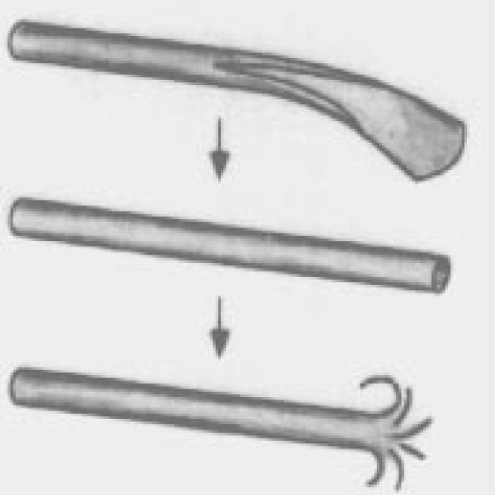
Fig. 2. According to the traditional concept of mesenchyme, during embryonic development, mesenchymal-like tissues form through EMT from epithelial-shaped tissues in two major steps at the early stages of gastrulation. Specifically, the primary mesenchyme originates from the epiblast to form mesoderm and primitive endoderm; this process is regarded as the prototype of EMT. Subsequently, the mesoderm reverts to an epithelial morphology to generate transient embryonic structures, including somites, which then undergo a further round of EMT to generate secondary mesenchyme. Secondary mesenchyme is considered the embryonic connective tissue and provides the precursors of the connective tissues of the adult body.
Mirco Galiè summarizes studies that support the hypothesis that the partially or fully mesenchymal phenotype might represent a general paradigm of stem cell plasticity underlying embryonic development, regenerative potential, as well as their pathological counterparts: https://bit.ly/3JADAW0
11.11.2025 17:14 — 👍 1 🔁 1 💬 0 📌 0
Interested in development? Cave fish? Enteric nervous system? Pretty pictures? Please join us next Friday! We are looking forward to the talk! 🤩
07.11.2025 21:48 — 👍 5 🔁 3 💬 0 📌 0
Biological Differentiation Across the Scales • November 14, 2025, at 12:00 p.m. Central • Register Now
One week left to register! Join the Editors-in-Chief of Differentiation on November 14 at 12:00 p.m. Central for a virtual seminar with Pavani P. Perera on the enteric nervous system, evolution, and cave fish. Register for free: https://bit.ly/4oaU71P
Topic: https://bit.ly/478UZhu
07.11.2025 19:49 — 👍 4 🔁 3 💬 0 📌 2

September/October cover of Differentiation. Fluorescence micrograph of Drosophila melanogaster testis tip. Merged testis image displays DAPI (yellow), anti-Mettl3 (cyan) and anti-Vasa (magenta) stains. Nuclear DAPI stain in combination with antibody against m6A methyltransferase Mettl3 and germline marker Vasa, indicates localization pattern of methyltransferase in cells of the apical tip during spermatogenesis.
Call for papers: Are you studying the role of splicing in development and how disruption of these processes leads to disease? Submit your manuscript to the “Splicing in Development and Disease” special issue by December 30, 2025! Learn more: https://bit.ly/4o6TAxW
04.11.2025 16:55 — 👍 1 🔁 0 💬 0 📌 0

Fig. 3. The most effective MSC exosomes-derived microRNAs applied for therapeutic approaches.
Review article: “Regenerative medicine and tissue engineering potential of mesenchymal stem cells exosomes-derived microRNAs,” by Navidreza Shayan et al. https://doi.org/10.1016/j.diff.2025.100911
28.10.2025 16:55 — 👍 1 🔁 0 💬 0 📌 0
Just announced: Join the Editors-in-Chief of Differentiation and Pavani P. Perera for the upcoming "Biological Differentiation Across the Scales" virtual seminar on November 14, at 12–1:30 p.m. Central!
Topic: https://bit.ly/478UZhu
Register for free: https://bit.ly/4oaU71P
27.10.2025 19:01 — 👍 1 🔁 1 💬 0 📌 0

Fig. 3. Effects of OPG on localization of CD44, CD47, DC-STAMP, ATP6V0D2 and Connexin43 in fusing OCs and precursors. From "Exploring the impact of osteoprotegerin on osteoclast and precursor fusion: Mechanisms and modulation by ATP," by Yunwen Peng et al.
NEW: The Editors-in-Chief of Differentiation, @rogerslabucd.bsky.social, @uribelab.bsky.social, and Loydie Jerome-Majewska, have curated a special collection of papers, “Osteo biology: from differentiation to regeneration.” Explore the collection: https://bit.ly/3JlXEey
22.10.2025 17:15 — 👍 7 🔁 7 💬 0 📌 0

Fig. 6. WNT5b mediates the Pi-induced suppression of OS cells migration. (A) Transwell assay was performed to evaluate the migration ability of NOS-10 (5 days) and SJSA-1 (10 days) cells with knock-down of WNT5b by siRNAs treated with osteogenic supplement medium. The analyses were performed at 24 h after seeding the cells into the culture inserts (n = 4). Scale bar = 500 μm.
Yuya Suzuki et al. evaluated the ability of two types of Pi (i.e., disodium phosphate [Na2HPO4] and monosodium phosphate [NaH2PO4]) to promote the osteogenic differentiation of osteosarcoma cells. Learn more: https://doi.org/10.1016/j.diff.2025.100912
21.10.2025 16:54 — 👍 1 🔁 0 💬 0 📌 0

Fig. 7. Atg13 knockdown in vivo enhanced the role of sirolimus in alleviating bone loss in OVX mice. OVX-operated female mice were treated with sirolimus (10 mg/kg, orally, 5 times/week) along with Cont-shRNA-AAVs or Atg13-shRNA-AAVs (5 × 10¹⁰ PFU/mL, marrow-cavity injection) for 45 days. (A) Representative 3D micro-CT reconstructed images of the tibiae from each group. Scale bar, 1 mm. (B) Representative H&E-stained tibial sections from each group. Scale bar, 20 μm. (C) LC3-puncta formation in bone marrow RANK+ CSF1R+ cells sorted by FACS was evaluated via immunofluorescence staining. Representative images of LC3 puncta, including single and merged fluorescence, were obtained under a fluorescent microscope. Scale bar, 25 μm.
“Inhibition of Atg13-mediated autophagy enhances the anti-osteoclastogenic effect of sirolimus by counteracting its pro-autophagic activity,” by Tingwei Gao et al.: doi.org/10.1016/j.di...
26.09.2025 13:38 — 👍 1 🔁 0 💬 0 📌 0

Call for Papers! Submit to the Splicing in Development and Disease special issue! Submission deadline: December 30, 2025. Guest editors: Natoya Peart and Karine Choquet
This special issue will address and highlight current understanding and gaps in knowledge about the role of splicing both constitutive and alternative in the development and how disruption of these processes leads to disease.
📢 Deadline extended: Submit to the "Splicing in Development and Disease" special issue by December 30, 2025!
Guest editors: Natoya Peart and Karine Choquet
Learn more and submit: https://www.sciencedirect.com/special-issue/322731/splicing-in-development-and-disease
24.09.2025 20:11 — 👍 0 🔁 0 💬 0 📌 0

Fig. 5. Osteogenesis imperfecta observed in X-rays of patients homozygous for WNT1 loss of function alleles. A) Limb fracture. B) Vertebral compression and deformation. C) Severe leg deformities.
Arne C. Lekven, Sarah Empie, and Richard Saoud review the history of Wnt1, its gene structure and regulation, expression, loss-of-function consequences, and connection to human disease. Read “One Wnt to lead them all: a Wnt1 primer” to learn more: doi.org/10.1016/j.di...
08.09.2025 21:05 — 👍 4 🔁 2 💬 0 📌 0
@rogerslabucd.bsky.social @uribelab.bsky.social @siegenthalerlab.bsky.social
23.06.2025 19:40 — 👍 1 🔁 0 💬 0 📌 0
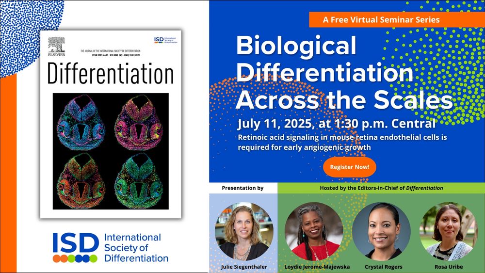
Biological Differentiation Across the Scales—July 11, 2025, at 1:30 p.m. Central—Retinoic acid signaliing in mouse retina endothelial cells is required for early angiogenic growth—Register Now!
Join the editors-in-chief of Differentiation on July 11 at 1:30 p.m. Central for a virtual seminar with Julie Siegenthaler on retinoic acid signaling in retina endothelial cells and early angiogenesis. 👁️🔬💉 Register for free: www.isdifferentiation.org/Events/Semin...
23.06.2025 19:39 — 👍 6 🔁 3 💬 1 📌 4
Cover of Differentiation
“The primary cilia: Orchestrating cranial neural crest cell development,” by Hiroyuki Yamaguchi et al., discusses the current understanding of the role of cilia and potential mechanisms of ciliogenesis in cranial neural crest cells. Learn more: Learn more: doi.org/10.1016/j.di...
19.05.2025 19:42 — 👍 1 🔁 0 💬 0 📌 0
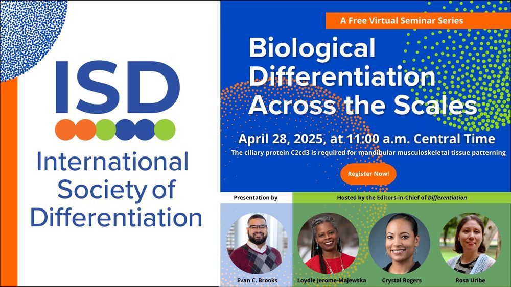
The ciliary protein C2cd3 is required for mandibular musculoskeletal tissue patterning. Evan C. Brooks, Ph.D. Graduate Student Alumnis, Brugmann Lab, Cinicinnati Children's Hospital Medical Center. Currently: Postdoctoral Fellow, University of Colorado School of Dental Medicine. "Biological Differentiation Across the Scales" Seminar, April 28, 2025.
The "Biological Differentiation Across the Scales" virtual seminar series kicked off on April 28 with an insightful, engaging talk by @ecbrooks96.bsky.social. Missed it live? Watch the recording: www.isdifferentiation.org/Journal/Page... @rogerslabucd.bsky.social @uribelab.bsky.social #devbio
06.05.2025 15:07 — 👍 4 🔁 7 💬 0 📌 0
This paper is now published at @diffjournal.bsky.social! Congratulations to all the hardworking students who made it happen! Shout out to authors 1 & 2! MJ is our “axolotl whisperer” and Raneesh is our computational wizard- both DVM/PhD students @ucdavisvetmed.bsky.social! 💫 doi.org/10.1016/j.di...
28.03.2025 03:22 — 👍 31 🔁 8 💬 2 📌 0
Excited to be the inaugural speaker for this new seminar series from @diffjournal.bsky.social. I’ll be presenting on work from my PhD thesis focused on the role of cilia in lower jaw development.
Thanks to Loydie Jerome-Majewska, @rogerslabucd.bsky.social, and @uribelab.bsky.social for the invite!
17.03.2025 20:19 — 👍 19 🔁 6 💬 1 📌 1
Join us for Biological Differentiation Across the Scales, a free virtual seminar series from Differentiation. Hear from authors and the editors-in-chief as they discuss key research. Stay at the forefront of biological differentiation—register now: www.isdifferentiation.org/Events/Semin... #devbio
17.03.2025 18:46 — 👍 8 🔁 3 💬 0 📌 3
Hello everyone! Differentiation has arrived on Bluesky! We are the flagship journal for the International Society of Differentiation with EICs- Loydie Jerome-Majewska, Crystal Rogers (@rogerslabucd.bsky.social), and Rosa Uribe @uribelab.bsky.social). Please consider us when you are submitting!
31.01.2025 21:40 — 👍 19 🔁 10 💬 0 📌 0
We publish the scientific journals @jcb.org, @jem.org, @jgp.org, @jhumimmunity.org, and co-publish @lsajournal.org. Visit our journals at https://rupress.org
Journal of Cell Biology publishes peer-reviewed research on all aspects of cellular structure and function. Published by Rockefeller University Press @rupress.org
🌐 https://rupress.org/jcb
Journal of Experimental Medicine publishes immunology, cancer, stem cells, microbial pathogenesis, vascular biology, and neurobiology research. Published by Rockefeller University Press @rupress.org
🌐 rupress.org/jem
Journal of General Physiology publishes research in physiological problems at cellular and molecular level - Published by Rockefeller University Press @rupress.org
🌐 rupress.org/jgp
Journal of Human Immunity (JHI) is the official open access journal of the International Alliance for Primary Immunodeficiency Societies (IAPIDS). Published by @rupress.org 🌐 jhumimmunity.org
#OpenAccess community-driven journal for peer-reviewed research in all areas of the life sciences. By EMBO Press (@embopress.org), Rockefeller University Press (@rupress.org), and Cold Spring Harbor Laboratory Press (@cshlpress.bsky.social).
Executive Editor at Life Science Alliance. I help decide how biology thinks 🤔
Views are my own and do not reflect editorial policy.
www.timfessenden.com
Deputy Editor at the Journal of Cell Biology
Views my own
Vive le Canada 🇨🇦
Cell Biologist at the University of Dundee, formerly at the MRC-LMB in Cambridge (UK) and Columbia University in NYC (USA). Interested in membrane traffic, protein quality control. Opinions my own. She/her.
Deputy Editor at Journal of Experimental Medicine, interested in host-pathogen interaction, innate immunity and beyond. Opinions are my own.
Senior editor at Journal of Experimental Medicine interested in all things immunology related and a soft spot for B cells. Opinions are my own.
We're a research lab at Rockefeller University (NY) and Necker Hospital (Paris) studying human genetic and immunological determinants of infectious diseases.
https://www.hgid.org/
#TCellsMakeYouSmart
This is my personal account where I will likely talk mostly about science… produced by us and others.
Pediatric Rheumatology & Immunology, #immunology, #immunodeficiency, #inbornerrorsofimmunity, #genetics, all opinions my own | https://megancooperlab.wustl.edu
We study the human immune system by examining host genetics. https://labs.vagelos.columbia.edu/bogunoviclab/
Pediatrician &
Professor of Pediatric Immunology
Human Systems Immunology
MRC LMS, Imperial College London
& Karolinska Institutet
Our research is driven to improve diagnosis and understand underlying mechanisms of inborn errors of immunity and autoinflammatory disorders.