We are recuiting two new Associate Professors here in Oxford Biochemistry. Come join us! Reach out to me if you have any questions. Please repost! tinyurl.com/mr3m7bd3
17.10.2025 14:56 — 👍 4 🔁 10 💬 0 📌 0
@matthew-batisio.bsky.social
Cellular Structural Biology Wellcome DPhil Student at The University of Oxford. Currently working in Peijun Zhang’s lab at The Oxford Division of Structural Biology. Passionate about biomedical animation and science communication: Batisio.co.uk
We are recuiting two new Associate Professors here in Oxford Biochemistry. Come join us! Reach out to me if you have any questions. Please repost! tinyurl.com/mr3m7bd3
17.10.2025 14:56 — 👍 4 🔁 10 💬 0 📌 0
SynapseNet is a deep learning based software tool that automates the segmentation of vesicles, mitochondria, synaptic compartments, and the active zone. This is visualized in three panels. On the left, a section of an electron tomogram with a synaptic compartment densely filled with vesicles, which appear as round structures with dark boundary and light body in the image, is shwon. The top right panel shows the segmentation result of vesicles, visualized by masks with an individual color per vesicle and outlines for the segmented compartment (red) and active zone (blue). The bottom right panels shows a 3D rendering of the segmentation with vesicles shown as yellow spheres.
Are you studying synapses in electron microscopy? Tired of annotating vesicles? We have the tool for you! SynapseNet implements automatic segmentation and analysis of vesicles and other synaptic structures and has now been published:
www.molbiolcell.org/doi/full/10....
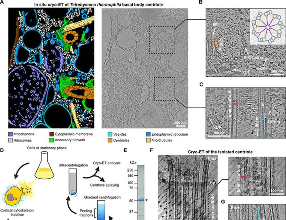
Huge congratulations to Wieczorek Lab PhD student Bin Cai and now-PI Jingwei Xu @xujwet.bsky.social on figuring out the surprisingly complex structure of the A-C linker crosslinking microtubule triplets in Tetrahymena basal body centrioles! www.science.org/doi/10.1126/...
10.10.2025 13:13 — 👍 22 🔁 8 💬 1 📌 1Happy to share the inaugural paper from the lab. We describe a molecular mechanism for the activation of outer dynein arm motors that power the vital motion of cilia.
Open access link below:
www.nature.com/articles/s41...
Here's a cool animated summary

New online: Molecular basis for the activation of outer dynein arms in cilia
29.09.2025 15:15 — 👍 10 🔁 3 💬 0 📌 1We have an open post-doc position in my group to study mRNA cleavage and polyadenylation using biochemical reconstitution and cryoEM.
Please get in touch if you are interested in joining this amazing team! 🔬🧬🤩
#RNA #cryoEM

Ensemble model of the full length Pex5 receptor (different blue color) in complex with Pex8 (green colors). Key Findings: Pex8 binds to a novel site on the mostly unfolded N-terminal region of the Pex5 receptor. This interaction is essential for peroxisomal protein import. Computational modelling reveals the formation of an assembly with the trimeric peroxisomal E3-ubiquitin ligase. We propose that this action positions the Pex5 receptor for its recycling, a step essential for the entire protein import process. Why this matters: Peroxisomes rely entirely on the import of folded proteins to function. Impaired peroxisome function is linked to severe disorders, and recent data show their crucial roles in carcinogenesis and the immune response. Our work elevates Pex8 from an enigma to a central player in this critical biological process. Read the full story and see the structures here: https://www.biorxiv.org/content/10.1101/2025.08.30.673231v1 #CellBiology #StructuralBiology #Peroxisome #ProteinImport #bioRxiv #Biochemistry
We are excited to share our latest work, where we unravel the structural and functional secrets of the once-mysterious protein Pex8 revealing how it controls the peroxisomal cargo import receptor Pex5.
Read more here: www.biorxiv.org/content/10.1101/2025.08.30.673231v1
Stunning cryo-ET from Peijun Zhang lab: Direct visualization of HIV-1 nuclear import!
Hundreds of viral cores captured entering the nucleus. The NPC dilates to let the capsid through. A masterclass in correlative microscopy that makes it quantitative. A leap for structural virology! @emboreports.org
Exciting to see our protein binder design pipeline BindCraft published in its final form in @Nature ! This has been an amazing collaborative effort with Lennart, Christian, @sokrypton.org, Bruno and many other amazing lab members and collaborators.
www.nature.com/articles/s41...
Want to acquire #ExM images like this and help us understand the true extent of cytoskeletal diversity across the tree of life? This position might be for you!
embl.wd103.myworkdayjobs.com/en-US/EMBL/j...
With @dudinlab.bsky.social
@embl.org @biology-unige.bsky.social @moorefound.bsky.social
🕒 NEW VIDEO 🕒
How can a cell know the time? Let's explore the beautiful 24-hour cycle produced by #KaiC proteins in the cyanobacteria S. Elongatus. Rendered in #blender3d with @bradyajohnston.bsky.social's brilliant Molecular Nodes add-on. #biology
Awesome video! I love how you represented the slow C1 domain hydrolysis.
06.08.2025 12:39 — 👍 0 🔁 0 💬 0 📌 0We are excited to announce the first in-cell structure of a LINC complex within a native nuclear envelope at subnanometer resolution!! #In-cell cryo-ET + #atomistic MD simulations! @tomdendooven.bsky.social et al!!
www.biorxiv.org/content/10.1...
Excited to share our latest work with @simonbullock11.bsky.social! We looked at how diverse mRNAs get selected for subcellular localization and it turns out that a single protein can recognize different RNA elements using shared features that weren’t apparent before.
www.biorxiv.org/content/10.1...
The Dunn School is a really great environment for taking up a Career Development Fellowship, and we are looking for candidates! Please spread the word.
31.07.2025 07:59 — 👍 6 🔁 4 💬 0 📌 0Very excited to announce that my first, first author paper, is now on BioRxiv! In this paper we unambiguously show #COPII coated vesicles in unperturbed human cells for the very first time!!
Make my day and check it out!
www.biorxiv.org/content/10.1...
#Cryo-ET #TeamTomo #Cryo-CLEM ❄️🔬❄️🔬❄️🔬❄️🔬
Thrilled to see our study on how kinesin-2 motors are switched on and off published in @natsmb.nature.com ⚛️
➡️ www.nature.com/articles/s41...
Congrats to all authors from me and Anthony 🎉 @dunnschool.bsky.social Check out this animation made by talented PhD student @matthew-batisio.bsky.social 😆
Less than two weeks left to apply!
Postdoc positions in my lab to study
1. initiation of DNA replication.
2. chromatin replication/epigenetic inheritance.
Great for biochemists, biophysicists & structural biologists.
Deadline 3 August 2025.
crick.wd3.myworkdayjobs.com/External/job...
Awesome work, and a beautiful graphical abstract!
18.07.2025 16:04 — 👍 1 🔁 0 💬 0 📌 0
Delighted to share the final version of Richard Zhou's study on how the CD163 scavenger receptor cleans up after toxic haemoglobin spills out from our blood cells. An amazing grabber mechanism!
rdcu.be/ewPA2

🎉 Excited to share my first paper as 2nd author! From the Barr & Gruneberg labs, this work explores the structure of the Chromosome Passenger Complex (CPC) and suggests a ‘pivot–tether’ model for how it binds H3pT3-phosphorylated nucleosomes
@oxfordbiochemistry.bsky.social @dunnschool.bsky.social
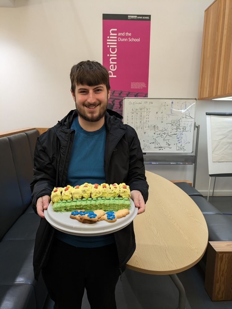
Matthew Clark holding a tray with a layered rectangular cake, showing the periodic repeating structure of IFT-A and IFT-B. Gingerbread cookies cut in the shape of dynein-2 ride on top.
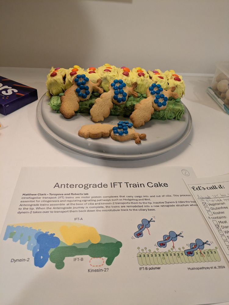
A close up photo of the cake on a table, showing the periodic repeating structure of IFT-A and IFT-B. Gingerbread cookies cut in the shape of dynein-2 ride on top.
IFT trains are really cool - and what better way to communicate their structure than baking a cake?
Created for my 1st year PhD project with Anthony Roberts and @kattoropova.bsky.social
IFT-A 🟡 IFT-B 🟢 Dynein-2 🔵
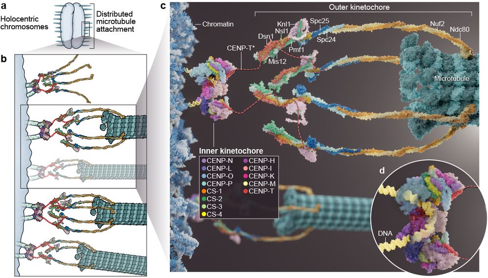
Happy to share our latest work on the structure and assembly of holocentric kinetochores! Huge thanks to Ines for a very fruitful collaboration, Claudio for all the support, and congratulations to Christine and all co-authors!
www.biorxiv.org/content/10.1...

Big news! 📢CCeMMP is holding the 2nd 'Bench to Art' Exhibition to showcase the artistic flair of structural biology. Share your creations to be displayed in a virtual gallery during National Science Week & for a chance to win cash prizes 💰! More info: ccemmp.org/events/arc-c...
#CryoEM
#Membranes

NEW pub: The role of metabolism in shaping #enzyme structures over 400 million years. Now out in @nature.com
Super grateful to have played a small role in this project - congrats to lead/corr authors Oliver, Benjamin, and Markus!
www.nature.com/articles/s41...
#alphafold #evolution #genomics
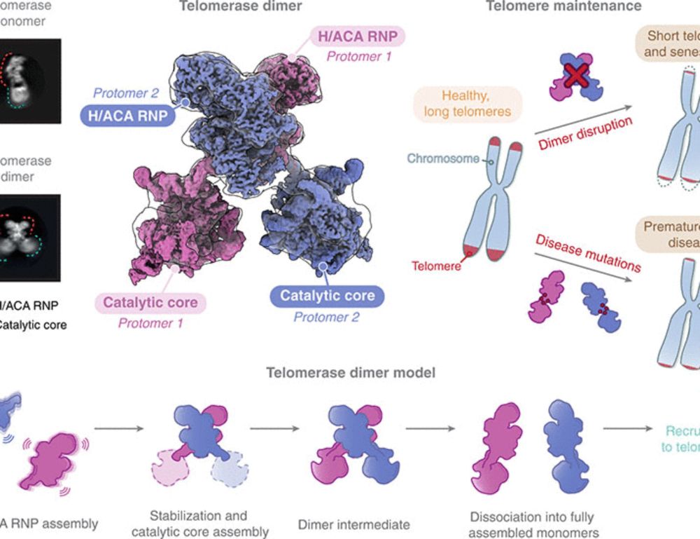
Proud to share our work where we resolved a longstanding question regarding the existence of a human telomerase dimer and provided insights into its function. Led by 3 amazing lab members in collaboration with @yiliangding.bsky.social and @rdaslab.bsky.social.
www.science.org/doi/10.1126/...
Excited to share our latest work! We found transcription factor SPT6 and phosphorylated Pol II CTD help recruit U1 snRNP to elongating Pol II, allowing efficient co-transcriptional splicing.
Glad to be featured in the Editors’ Highlights!
www.nature.com/articles/s41...
#splicing #cryoEM
New Independent Fellowship position in Microbiology to launch your lab in our department @johninnescentre.bsky.social (UK). We are conducting a broad search in the area of plant-associated microbial interactions. Message me if you have any questions.
Apply here: www.jic.ac.uk/vacancies/in...
Brilliant animation of an incredibly gymnastic process! I remember going to a John Diffley lecture in 2023 and loving this mechanism.
02.07.2025 19:17 — 👍 1 🔁 0 💬 0 📌 0🦠 NEW VIDEO 🦠
The molecular biology of how Mycobacterium Tuberculosis evades the immune system so well.
Starting with this simulation of a full outer envelope of m.TB--let's explore how the deadliest & most persistent human pathogen completely hacks our immune response. #molecularbiology