Deep Learning for Microscopy Image Analysis
Topics The following will be covered extensively during lectures, exercises, and project work: Image denoising and restoration (fully supervised and self-supervised) Image translation (e.g.,
🚨 Alarm!!! 🚨
AI/ML course for microscopy image analysis!!! 🧐
In 2026 at Janelia (@hhmijanelia.bsky.social), no tuition, housing and meals provided! Isn’t that borderline unbelievable?!?
20 students, ~14 TAs and lecturers
🗓️ June 4-18 2026
✍️ Jan 15 2026 ✍️
🔁 pls!!
www.janelia.org/you-janelia/...
28.12.2025 19:05 — 👍 78 🔁 62 💬 0 📌 3

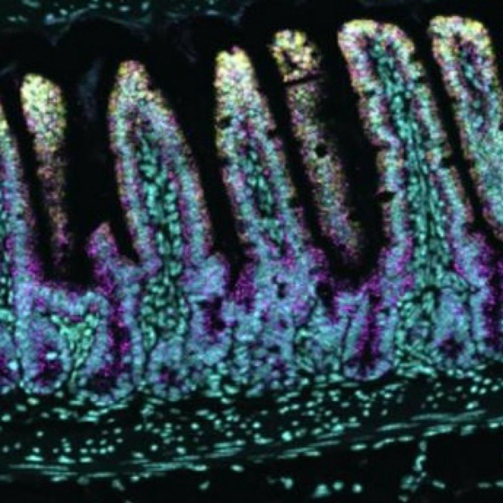
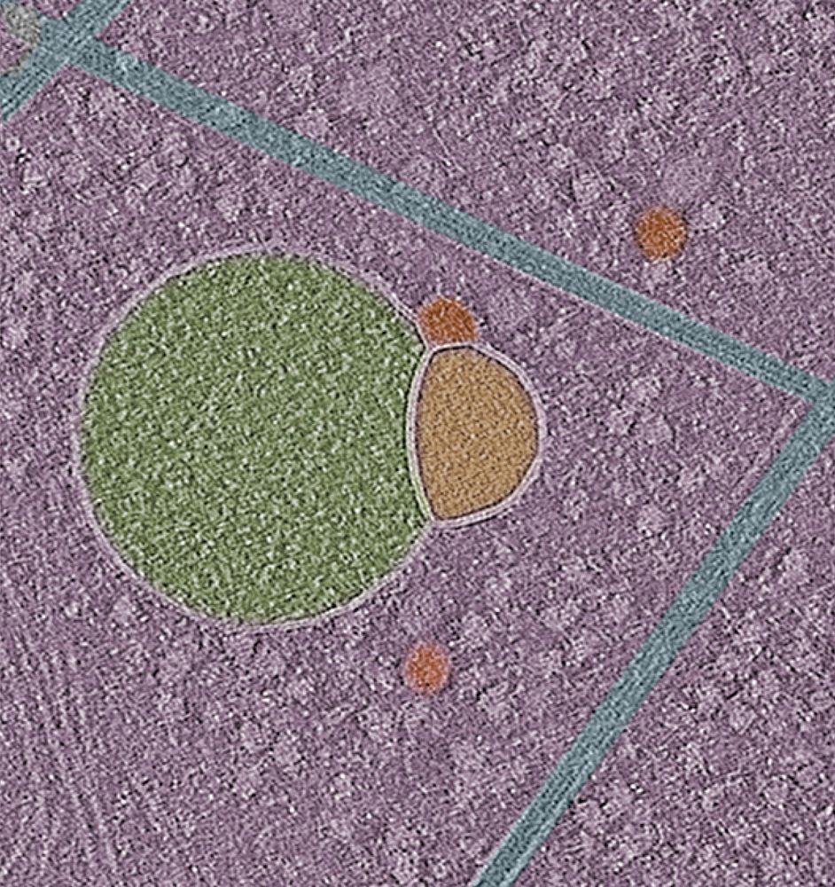
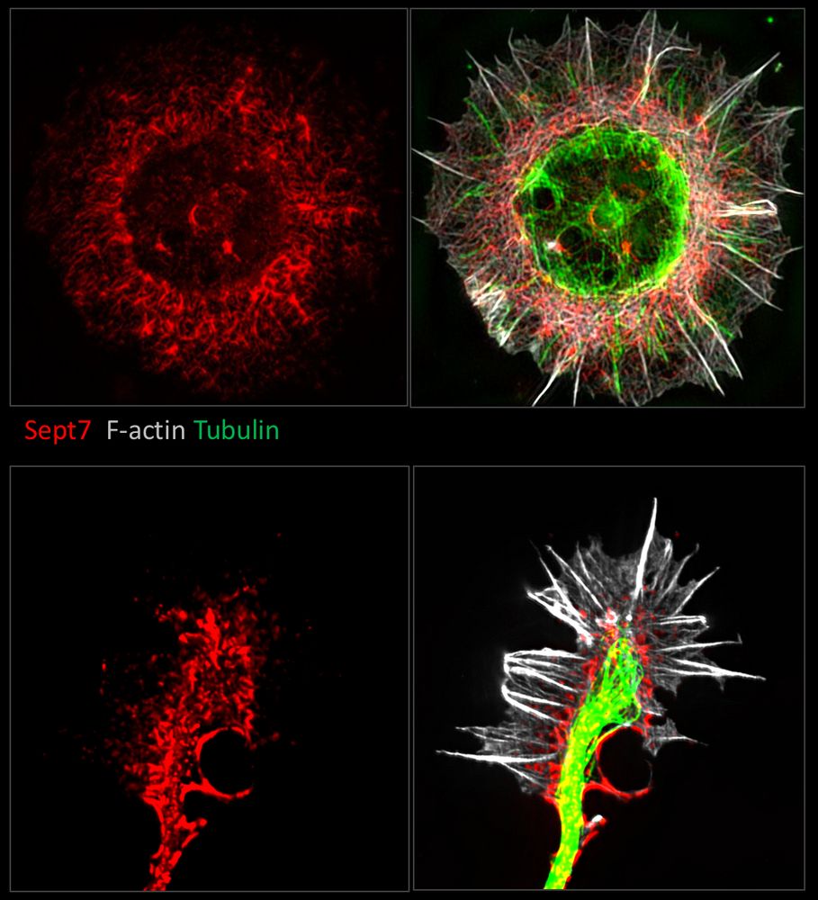
From earlier this week: This issue’s Cover: @ebrahim-lab.bsky.social @dominikrobak.bsky.social & team report on the septin cytoskeleton at apical junctions of intestinal epithelial cells, whereby it maintains barrier integrity & protects from inflammation: insight.jci.org/articles/vie...
29.11.2025 14:01 — 👍 5 🔁 2 💬 0 📌 0
Thanks, Helge!!
25.11.2025 11:35 — 👍 1 🔁 0 💬 0 📌 0
Very excited to share the latest work from our lab in @insight.jci.org, highlighting a role for the #septin #cytoskeleton in protecting against "leaky gut", by recruiting non-muscle myosin II and stabilizing the tight junction.
Credit to @dominikrobak.bsky.social for the stunning cover image!
24.11.2025 18:01 — 👍 17 🔁 8 💬 1 📌 2
Check out our new preprint on the discovery of a molecular switch in NAC that mediates nascent chain sorting on the ribosome and prevents mitochondrial protein mistargeting by SRP. A great collaboration with the Shan Lab @Caltech and the Qi Lab @UVA: www.biorxiv.org/content/10.1...
01.08.2025 15:27 — 👍 82 🔁 28 💬 4 📌 2
Having seen your bingo card, we had to make it happen 😉!
Thank you for highlighting our work @hankgreen.bsky.social ! 🙏🏽
26.07.2025 01:17 — 👍 2 🔁 0 💬 0 📌 0
Thank you, Ilya! :)
19.05.2025 18:12 — 👍 1 🔁 0 💬 0 📌 0

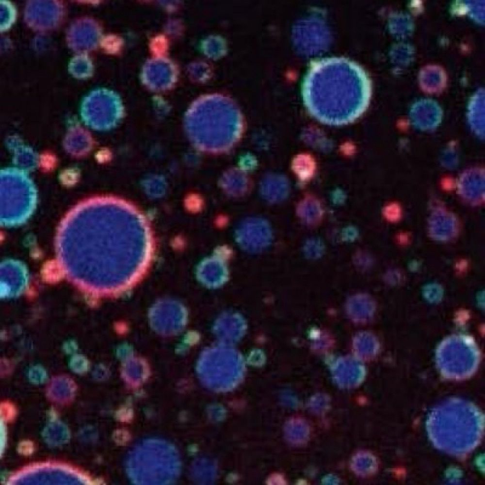
Proposed mechanism of MVB formation
17.05.2025 20:46 — 👍 1 🔁 0 💬 0 📌 0
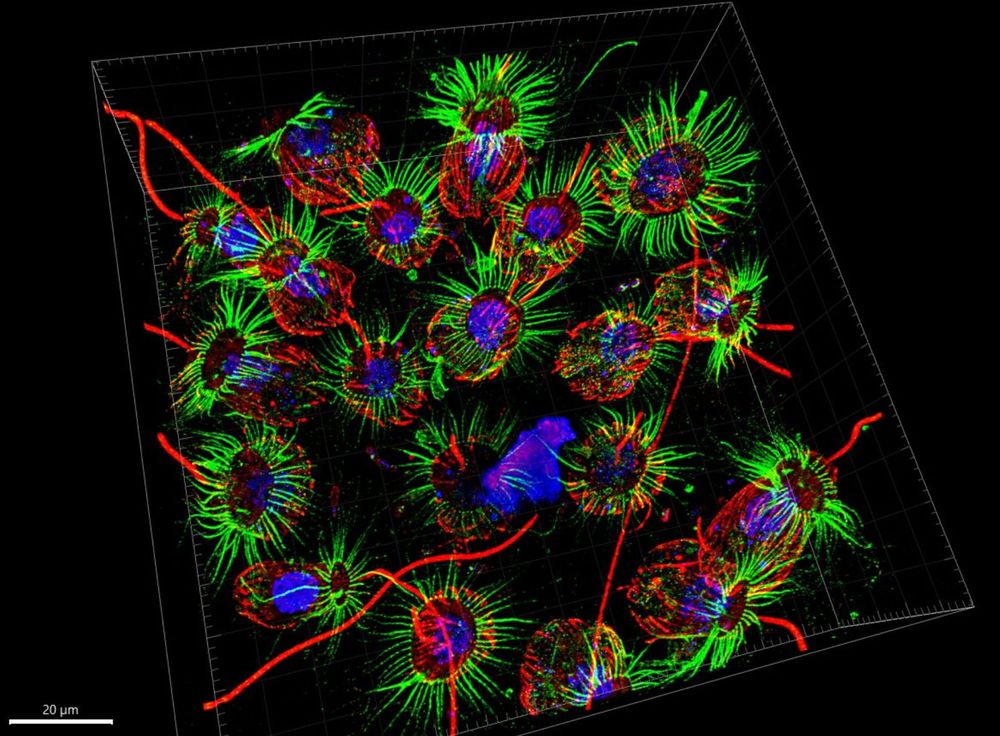
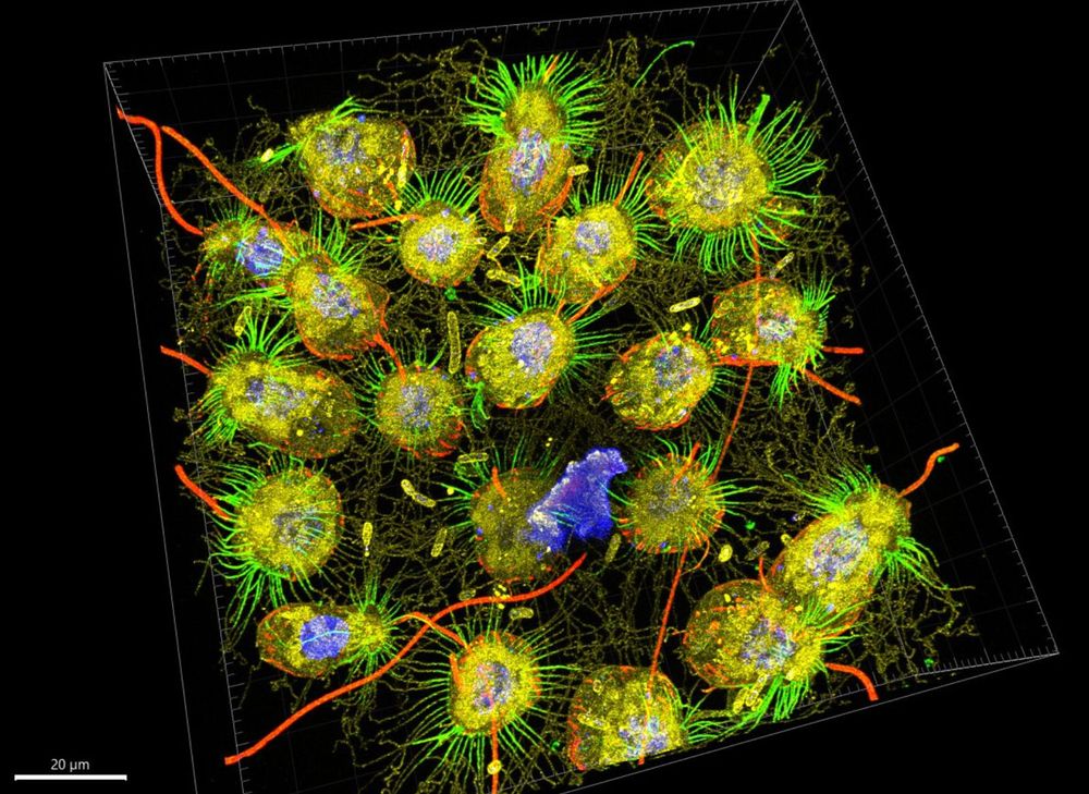
Early for #MicroscopyMonday: #Hemifusomes- Newly discovered ESCRT-independent intermediates of intraluminal vesicle and multivesicular body formation! In collaboration with the Kachar Lab at the NIH and out today in @natcomms.nature.com : rdcu.be/emtQu
#cryoET #trafficking #organelle #hemifusion
17.05.2025 19:37 — 👍 36 🔁 9 💬 3 📌 2
😞
06.05.2025 21:44 — 👍 0 🔁 0 💬 0 📌 0

"Stereocilia"- actin-based skyscrapers that adorn sensory cells of our inner ear and covert vibrations into electrical signals—the first step in hearing and balance. #FluorescenceFriday
02.05.2025 13:58 — 👍 20 🔁 4 💬 0 📌 0
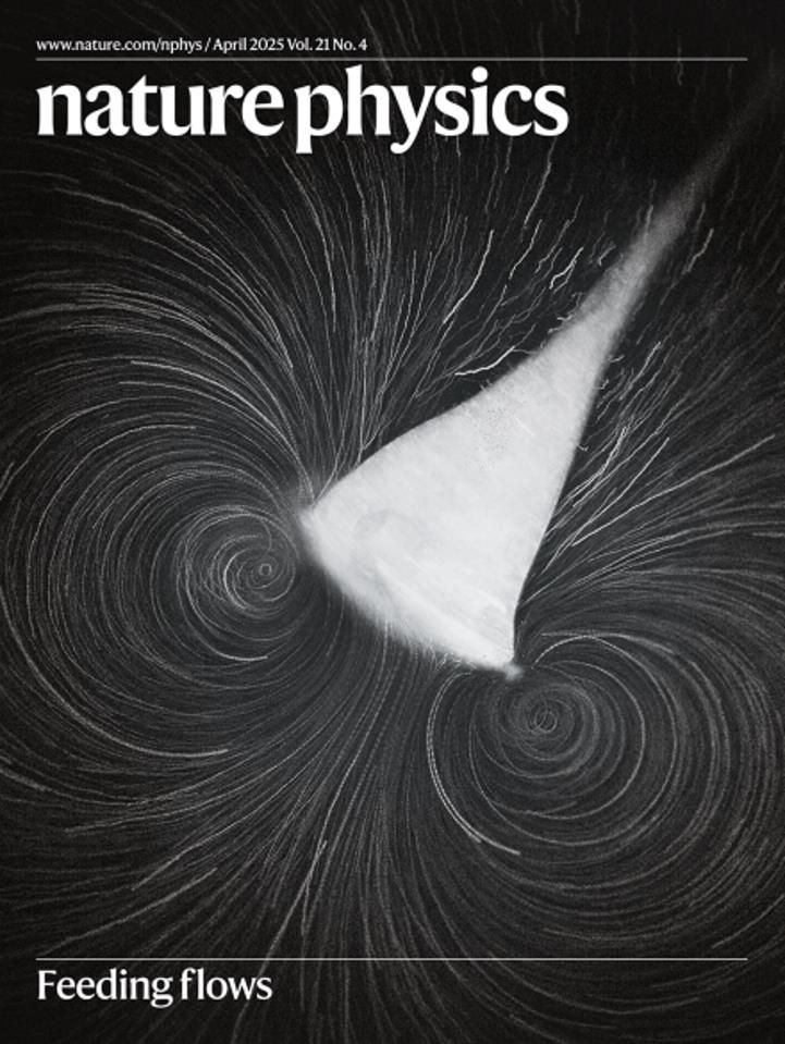
🎉🎉We’re on the cover of Nature Physics! @natphys.nature.com
Here is the original paper www.nature.com/articles/s41...
26.04.2025 14:38 — 👍 48 🔁 8 💬 1 📌 0

From stem cell to structure-
This human colonoid shows the self-organization of intestinal epithelial cells in 3D—nuclei in magenta, actin in green. A living miniature gut for your #FluorescenceFriday.
25.04.2025 11:07 — 👍 18 🔁 1 💬 0 📌 0
Expansion microscopy micrograph of the choanoflagellate Choanoeca flexa, vaguely evocative of a cactus. Red = tubulin, green = actin, blue = DNA
Same with yellow = lipids (BODIPY)
A dream come true: the first expansion microscopy images of C. flexa 🤩 Generated by Mylan & Uzuki who learned from the best (@hiralshah.bsky.social @gautamdey.bsky.social @dudinlab.bsky.social). We will learn so much from these!
22.04.2025 16:36 — 👍 293 🔁 58 💬 20 📌 9
Only 29 slots left out of 200--register now!
21.04.2025 14:12 — 👍 8 🔁 7 💬 0 📌 0

🧫 A gentle reminder that your gut is an actual work of art.
#FluorescenceFriday #BeneathTheScope
18.04.2025 14:33 — 👍 36 🔁 7 💬 0 📌 0
Two months left for FASEB Conference - Septins: Spatial regulators of cell biology
Register now:
events.faseb.org/event/septin...
Come find out what these filaments do at the interface of the actin and microtubule cytoskeletons and cell membranes. You'll be hooked for life.
16.04.2025 15:08 — 👍 11 🔁 5 💬 0 📌 1
Is liver zonation conserved from mice to humans? And how does fibrosis affect zonation in patients? Now, these questions are answered using scDVP. Some collaborations are meant to be! @carolineweiss.bsky.social@frosenberger.bsky.social @mannlab.bsky.social@mlsb-borgwardt.bsky.social
14.04.2025 14:35 — 👍 8 🔁 6 💬 0 📌 0


#MicroscopyMonday | Colon-scoping turned coral reef
Was imaging the colon when I accidentally stumbled into this glowing patch of vascularized white adipose tissue.
🔵 mT/mG mouse (mT-only) → red membranes shown in cyan
Accidental imaging hits different.
#IntravitalImaging #LiveImaging
15.04.2025 00:01 — 👍 2 🔁 1 💬 1 📌 0
Excited to share our preprint on the molecular architecture of heterochromatin in human cells 🧬🔬w/ @jpkreysing.bsky.social, @johannesbetz.bsky.social,
@marinalusic.bsky.social, Turoňová lab, @hummerlab.bsky.social @becklab.bsky.social @mpibp.bsky.social
🔗 Preprint here tinyurl.com/3a74uanv
11.04.2025 08:35 — 👍 359 🔁 141 💬 12 📌 20
#3DThursday | Intestinal villi in all their folded glory
These self-renewing, nutrient-absorbing structures are beautiful and functional.
🔴 Phalloidin
🔵 DAPI
🟢 Clvd cas-3 (extrusion in action!)
Fixed tissue 3D imaging = big insight, tiny resolution.
#Microscopy #3DImaging #biophysics #intestine
11.04.2025 03:08 — 👍 2 🔁 1 💬 0 📌 0
Molecular Biology of the Cell (MBoC)
Have cutting-edge cell biology research to share? Submit to Molecular Biology of the Cell (MBoC)- dedicated to advancing discovery and fostering collaboration.
Proud to be part of the editorial team supporting groundbreaking science. molbiolcell.org
#CellBiology #MBoC #Research
08.04.2025 19:15 — 👍 2 🔁 0 💬 0 📌 0
Deputy Editor Science magazine
Areas of responsibility include: Cell biology, cellular microbiology, membrane traffic, protein targeting, protein folding, organelle biogenesis, cytoskeleton, cell polarity, prion biology, developmental biology, stem cells
Associate professor at UCSF. Cell biologist at heart, research focus on the function and dynamic regulation of the nuclear periphery.
www.buchwalterlab.UCSF.edu.
Whitehead Institute and Department of Biology, MIT. Lover of cell biology and cell division. Aspiring to do good science and do good.
A scientific journal publishing cutting-edge methods, tools, analyses, resources, reviews, news and commentary, supporting life sciences research. Posts by the editors.
We study antiviral immune responses of the intestinal epithelium, especially interferon lambda. We focus on how they are specialized for the gut, interactions with normal microbiota at homeostasis, and how they may go awry during inflammatory diseases.
Researcher at the Centre for Military Studies, in Copenhagen. Working on ethics, politics, technology, Just War, drones, and autonomous weapons.
PhD student at Karolinska Institutet and Scilifelab | Cell physics
https://www.csi-nano.org/
Post-doc at CU Boulder #sko | cell and cancer biology | 🔬 and 🔭 content welcome
Assistant Professor, The Pennsylvania State University, Department of Cell and Biological Systems, Hershey, PA
(Re)searcher of non equilibrium steady states living at the interface of physics and biology. Leading the Living Patterns lab @EPFL
https://www.epfl.ch/labs/lpl/
Mechanobiologist-turned-organoid architect | Cell-cell adhesion afficionado | Imaging as coping mechanism.
PhD-ed from @mbisg.bsky.social
Postdoc at Koehler Lab
@Bostonchildrens.bsky.social
@Harvardmed.bsky.social
High-quality, topflight, and clinically impactful basic and translational research representative of all biomedical specialties and physician-scientists: https://insight.jci.org/
Physiologist, ERAD biology, Professor and Chair, Dept of Molecular Physiology and Biological Physics @UVA; formerly at U-Michigan and Cornell University
@CNRS Research Director @ENS_ULM. Interested in Droplets, Metabolism, Organelles, Lipids, BioPhysics, Microfluidics & Co, 🇸🇳
Professor, Oakland University, MICHIGAN.
Visiting Faculty@ LSI, UMich (Klionsky lab).
| Chromatin |Transcription |Autophagy | Yeast | Genomics |🧪
Assistant Professor @OhioState in a toxic relationship with lysosomes. ECR Editor @MBoCjournal. Former @NIH. Views are my own. http://bonetponcelab.com/
Researcher @UCSF working on the presynaptic dysregulation in #Parkinson's disease 🧠🔬 avid reader 📚 coffee addict ☕️ | Previously @EdinburghUni
Studying mitochondrial responses to infection at UCLA | Mitochondriac | Autophagy| DEI | Outdoor enthusiast | he/él | 🇲🇽⛰🚴🏼🏳️🌈
https://josedelgadophd.wixsite.com/jose
Uncovering macrophage cell biology using whole-genome screens and microscopy. My lab studies regulation of phosphoinositides and the actin cytoskeleton in the context of macropinocytosis, as well as lipid droplet formation and metabolism.