We are moving - new place, new ideas...
07.10.2025 07:07 — 👍 1 🔁 0 💬 0 📌 0
@vweinhardt.bsky.social
physicist developing 3D and 4D bioimaging techniques particularly X-ray tomography across multiple resolution and contrast scales


Pan-ASLM system for large scale expansion microscopy from the Bewersdorf lab.
www.biorxiv.org/content/10.1...
Repost appreciated:
Open #PhDposition in structural virology in my group! Join us in Umeå, Sweden to uncover how arboviruses remodel infected cells. In situ #cryoET and #cryoEM combined with #virology, #cellbiology, and #biophysics.
Deadline 7 September. More info: www.carlsonlab.se/join/

The figure shows principles of local tomography and alignment strategy for sparse and high-resolution projection images. At the bottom of it, there are three images with the results of the interior scan, full view, and combined reconstructions.
🚀 Our latest preprint introduces a sparse global sampling approach for interior SXT.
This enables:
🔍 “Scout & zoom” imaging of organelles in 3D
🎯 Optimized radiation dose without losing resolution
📊 Quantitative absorption maps from sparse data
📄 Read more: www.biorxiv.org/content/10.1...

🎙️ I am excited to announce that our latest progress on automatic segmentation of cell anatomy has been published in the IEEE!
🔬 Segmentation of cellular organelles is one of the main challenges in SXT.
#softxraytomography #cellbiology #DeepLearning #IEEE
👓 Read the full article here:

The Weberian apparatus boosts hearing in otophysan fish via modified bones and ligaments. Using advanced imaging and histology, a study from @jakeleyhr.bsky.social and
@remorphevolab.bsky.social reveals new insights into the elusive first intercostal ligament and its role in this acoustic system
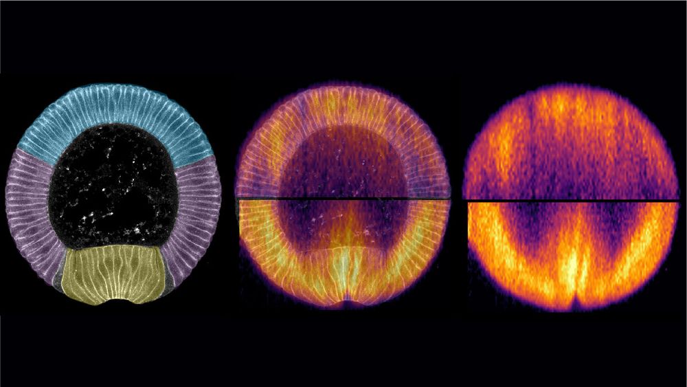
🥁 New article 📢: #Mechanobiology of #development during #Drosophila #gastrulation using #Brillouin microscopy, now in @natcomms.nature.com : rdcu.be/ev6ZX
Collab. w/ @Prevedel_Lab @embl.org , Maria Leptin @marialep.bsky.social, @abhisha-thayambath.bsky.social, Julio Belmonte @ncstate.bsky.social
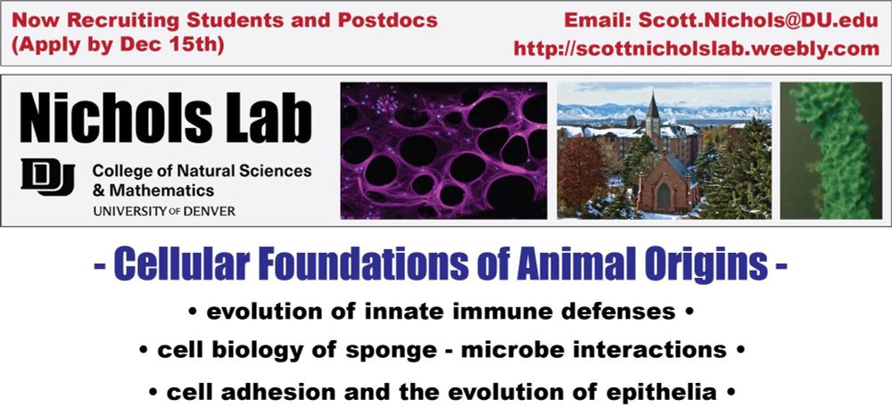
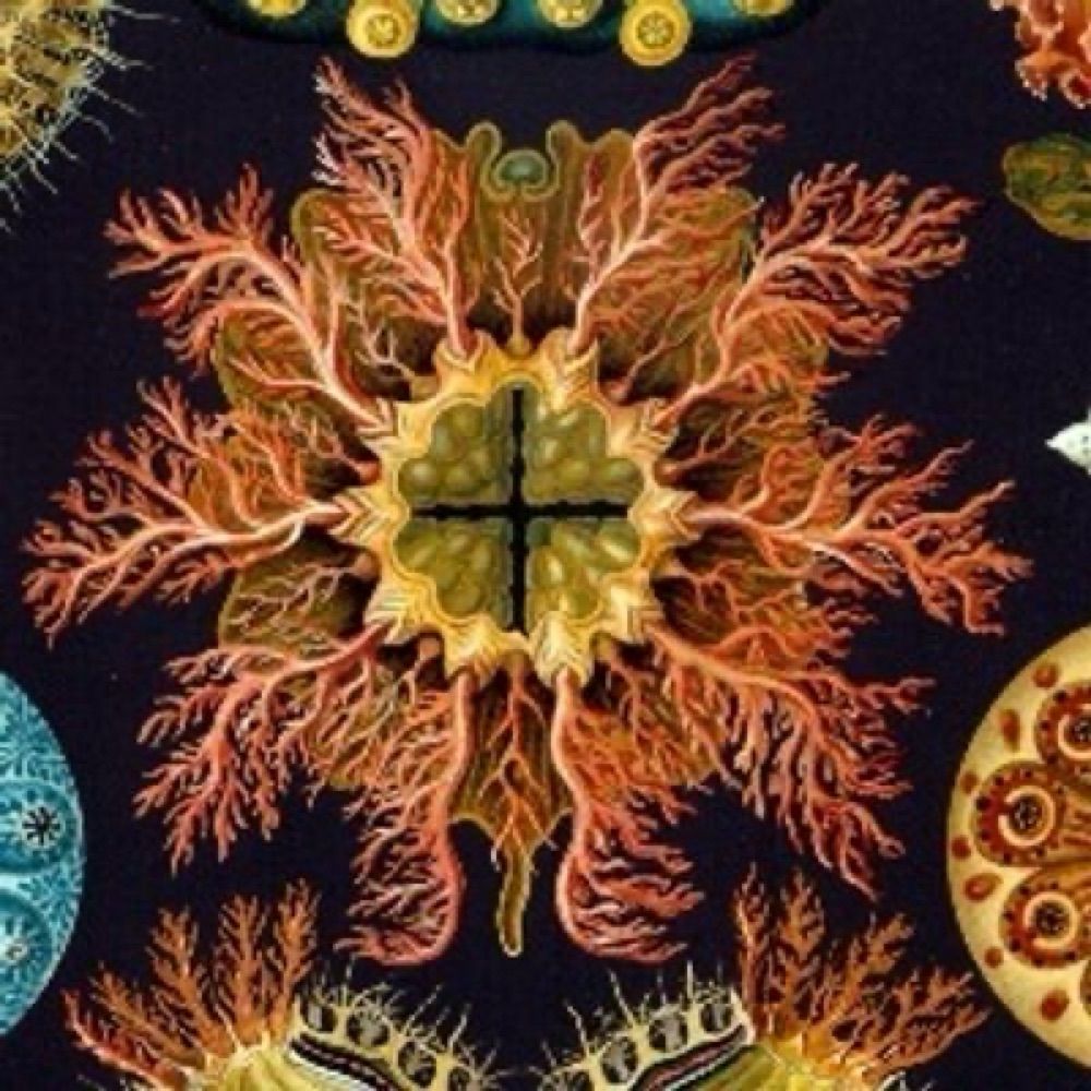
Please help get the word out - We are hiring at all levels (graduate students, technicians, and postdocs)! If you are interested in sponges, cell biology, evolution, immunity, and symbiosis, please don’t hesitate to contact me to learn more.
16.07.2025 19:36 — 👍 12 🔁 16 💬 0 📌 0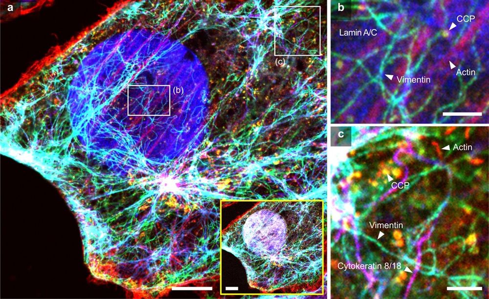
Registration of multiplexed images via actin staining in expanded cultured cells. (a) Composite image generated by registering two images acquired from the first and second imaging rounds. Phalloidin-labeled actin fibers were used as fiducial markers. The yellow box displays six-color images, including the DAPI channel. Gray, nucleus; red, actin; cyan, vimentin; blue, laminA/C; yellow, CCP; and magenta, cytokeratin 8/18. (b–c) Magnified views of the boxed region in a. Scale bars: (a) 2 µm; (b–c) 500 nm.
#ExpansionMicroscopy allows multiple rounds of staining & imaging, but requires high-quality registration of images between rounds. This study develops a registration technique using dense NHS-ester labels for multiplexed cyclic imaging in expandable tissue gels @plosbiology.org 🧪 plos.io/44M4wcX
04.07.2025 12:53 — 👍 21 🔁 8 💬 0 📌 0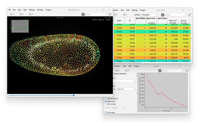
We just released and uploaded a new version of *Mastodon*, the large-scale tracking and track-editing framework for large, multi-view images.
Mastodon is available via Fiji, via a dedicated update site.
mastodon.readthedocs.io/en/latest/do...
Below is a thread that survey some of the novelties ⏬️

🚀 Thrilled to be named "Physikerin der Woche" by #DPG! A celebration of women shaping physics—proud to join this inspiring group. Dive into stories of science and resilience! #PhysikerinDerWoche #WomenInPhysics
www.dpg-physik.de/vereinigunge...
Congratulations! Amazing to see this cluster succeeding
22.05.2025 21:53 — 👍 1 🔁 0 💬 0 📌 0Further attempts to stay positive and push the boundaries of X-ray imaging in plant biology, here's a maize root tip. Technical voxel resolution is 0.8um, field of view is about 1mm tall, using the 20X lens on my ZEISS Versa 520 XRM.
@danforthcenter.bsky.social
@zeiss-microscopy.bsky.social
🎉 Congratulations to Manisha Biswas for winning the #SocialSciences category in this year's #DanceYourPhD competition.
Watch the entire video on the power of collective synchrony—and learn more about the other category winners: scim.ag/4lSpjCl
🚀🔬🦠 Releasing 🤖Cellpose-SAM🤖, a cellular segmentation algorithm with superhuman generalization 🦸♀️. Try it now on 🤗 huggingface.co/spaces/mouse...
paper: www.biorxiv.org/content/10.1...
w/ @computingnature.bsky.social 1/n

Spatial mechano-transcriptomics👹
Image-based cell mechanics inference
Segmentation Multi-junctional molecules▶️
Circular arc polygon tiling for cell contacts▶️
Cellular pressure, junctional tension, stress tensor
+Imaging-based #SpatialTranscriptomics
#NatMethods 2025
www.nature.com/articles/s41...
🚨We have a fully funded open PhD position in bacterial synthetic biology available in our lab (www.yschaerli.com) in beautiful Lausanne, Switzerland @dmf-unil.bsky.social @fbm-unil.bsky.social @unil.bsky.social
If you are passionate about #SynBio and #GeneCircuits, apply here: tinyurl.com/mry2xwa5
📢 Preprint alert 📢
(1/6) Our work on 3D force inference for intestinal organoids is now on bioRxiv:
www.biorxiv.org/content/10.1...
Stunning collaboration between @omdrozdowski.bsky.social and @kimboonekamp.bsky.social from the lab of @michaelboutros.bsky.social and with Ulrike Engel. A short🧵...

Calling all #MedakaPI researchers! Join us in Heidelberg, Germany from July 22-24, 2025 for our collaborative meeting! Register now & take advantage of early bird rates until April 15th! meetmedaka.com/registration/ 🐟 #MedakaResearch #ModelOrganisms #ScienceConference #FishModels #Heidelberg2025
07.04.2025 13:31 — 👍 8 🔁 6 💬 0 📌 2
We are excited to share that our NanoX project has been featured in the Rhein-Neckar-Zeitung @rnzonline.bsky.social! Thank you to Amira Sanli for visiting our laboratories and showcasing our work in the newspaper!
#NanoX #HepatitisE #softxraytomography #cellbiology
We recently described the Axillary Lymphoid Organ (ALO) in the #Zebrafish. It's on the OUTSIDE of the fish, making it great for imaging! Learn more by checking out our paper in @jem.org rupress.org/jem/article-...
03.04.2025 00:07 — 👍 60 🔁 21 💬 2 📌 1
Fantastic piece on the power and gravity of research that on it face may seem trivial. @labonnelab.bsky.social reminds us that basic research is the foundation of ALL research.
www.statnews.com/2025/04/03/b...

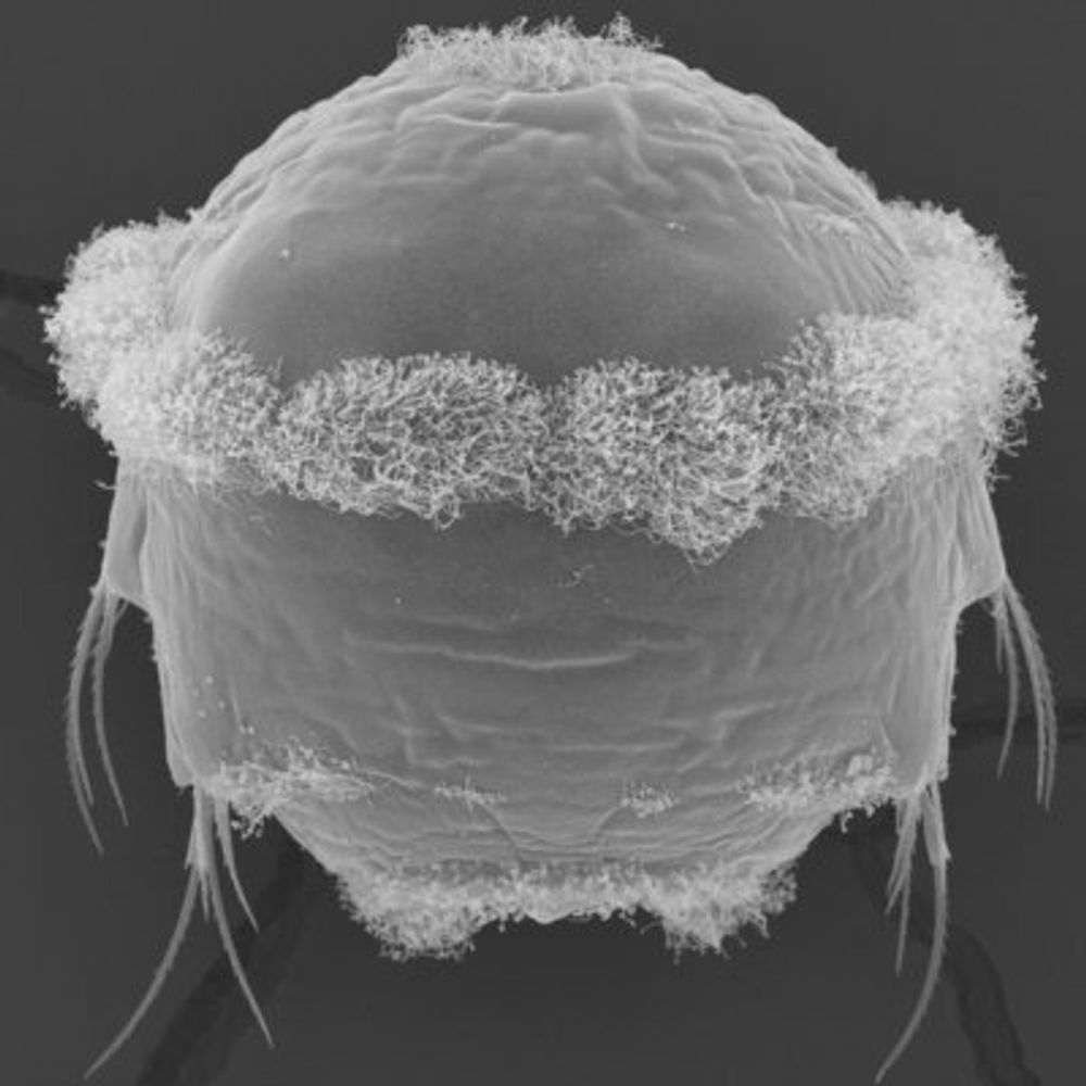
eft: A 2D CryoSXT projection of an HSV-1 evelopment event. Right: A 3D rendering of the budding assembly stage captured by CLXT.
A new combination of microscopy methods has revealed exquisite details of the virus assembly process used by herpes simplex virus during replication.
buff.ly/aO8W9BJ

My presentation on "Coordination Across Scales — Or Why We Study Organisms"
at the "Future 3D Additive Manufacturing – The 3DMM2O Conference" from today.
https://jekelylab.github.io/3DMM2O_Kloster_Schoental_Mar_2025.html#/title-slide
#cilia #neuroscience #larva #biology

Come check out poster 185 in tonight's session at #biologists100 🐣! And for more detail, here's the full article recently published in @plosbiology.org
journals.plos.org/plosbiology/...
New observations on white matter organisation at the micron scale. #Neuroscience
elifesciences.org/articles/949...

Our review on Soft X-ray Tomography (SXT) got just published in Annual Reviews!
🔬 We covered everything from the tech to the software to the latest discoveries.
Read the full review 👉 tinyurl.com/379juxbn
#softxraytomography #cellbio #microscopy #imaging #science #research #AnnualReviews

Press release on the self-driving multiscale microscope from @daetwylerstephan.bsky.social et al:
www.utsouthwestern.edu/newsroom/art...

A study by Viktória Parobková from CEITEC BUT and co-authors presents the use of micro-CT to image microplastics without damaging the biological sample. The study demonstrates the benefits of the methodology on zebrafish (Danio rerio) 🐠
Read and see micro-CT scans ⬇️
www.ceitec.eu/scanning-zeb...
eft: A 2D CryoSXT projection of an HSV-1 evelopment event. Right: A 3D rendering of the budding assembly stage captured by CLXT.
A new combination of microscopy methods has revealed exquisite detail of the virus assembly process used by herpes simplex virus during replication.
buff.ly/aO8W9BJ