
 31.12.2025 23:25 —
👍 1
🔁 0
💬 0
📌 0
31.12.2025 23:25 —
👍 1
🔁 0
💬 0
📌 0
31.12.2025 23:25 —
👍 1
🔁 0
💬 0
📌 0
31.12.2025 23:25 —
👍 1
🔁 0
💬 0
📌 0

Best wishes for 2026 to everyone from our Lab at
ebrahimkhanilab.bio
We look forward to a year defined by discovery, curiosity, and bold exploration.
Stay tuned for more news coming soon!
#organogenesis #stemcellmodels #syntheticbiology #howtissuesarebuilt
🤩 Very excited to share our new work! We have derived euploid and aneuploid trophoblast organoids and extra-embryonic mesoderm cell lines from early human embryos. In doing so, we have characterised the tissue requirements for their specification. If you want to know more, continue reading….
09.08.2025 08:03 — 👍 37 🔁 11 💬 1 📌 0
A big thanks to all authors, especially Professor Naihe Jing and Penglei Shen for guiding our embryo model into organogenesis, and to our PhD students Jiahui Huang and Wei Guan for their efforts. And of course, to Huanhuan Li, the driving force behind this work.( 3/3)
07.08.2025 15:58 — 👍 2 🔁 1 💬 1 📌 0
Starting from PSCs, a brief reprogramming step gives rise to “embryo founder-like cells” (EFCs). EFCs segregate embryonic and extraembryonic lineages and self-organize to closely mirror natural embryo development, both morphologically and at the molecular level.
authors.elsevier.com/sd/article/S...
Very happy to share the work of Huanhuan Li et al. While not a natural embryo, you might not have known that if I hadn’t told you. (1/3)
07.08.2025 15:58 — 👍 14 🔁 8 💬 3 📌 0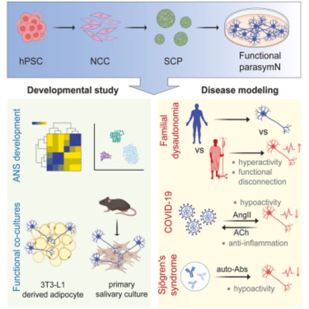
Research highlight from Cell Stem Cell: Parasympathetic neurons derived from human pluripotent stem cells model human diseases and development
www.cell.com/cell-stem-ce...
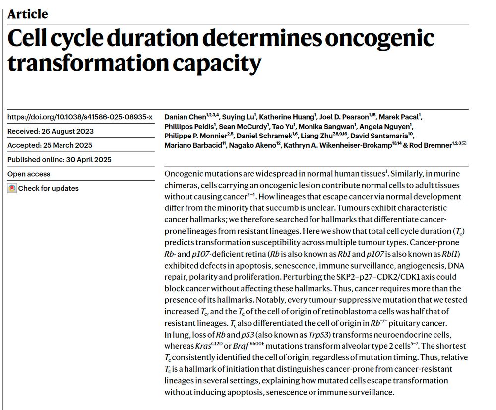
Cell cycle duration determines oncogenic transformation capacity Danian Chen, Suying Lu, Katherine Huang, Joel D. Pearson, Marek Pacal, Phillipos Peidis, Sean McCurdy, Tao Yu, Monika Sangwan, Angela Nguyen, Philippe P. Monnier, Daniel Schramek, Liang Zhu, David Santamaria, Mariano Barbacid, Nagako Akeno, Kathryn A. Wikenheiser-Brokamp & Rod Bremner https://www.nature.com/articles/s41586-025-08935-x
A recent paper reveals a surprising insight. The length of the cell cycle in a particular tissue determines the oncogenic transformation capacity of particular oncogenes, with tissues with shorter cell cycles most susceptible 3/n
26.07.2025 14:13 — 👍 57 🔁 21 💬 4 📌 2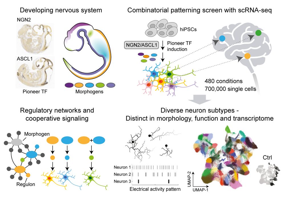
Neuron programming! Pro-neural TFs + 480 morphogen conditions + scRNA-seq --> Diverse iN subtypes of forebrain, midbrain, hindbrain, spinal cord, and PNS. @hsiuchuanlin.bsky.social @jasperjanssens.bsky.social and Treutlein Lab! @science.org www.science.org/doi/10.1126/... #NGN2 #ASCL1
11.07.2025 20:59 — 👍 78 🔁 32 💬 4 📌 0
New preprint: How do cells measure time? In human development, pluripotent epiblast cells pause for about two weeks before differentiating into more specialised cell types. We show that this timing is controlled by a transcriptional clock that works like an hourglass. www.biorxiv.org/content/10.1...
19.03.2025 11:29 — 👍 10 🔁 3 💬 1 📌 0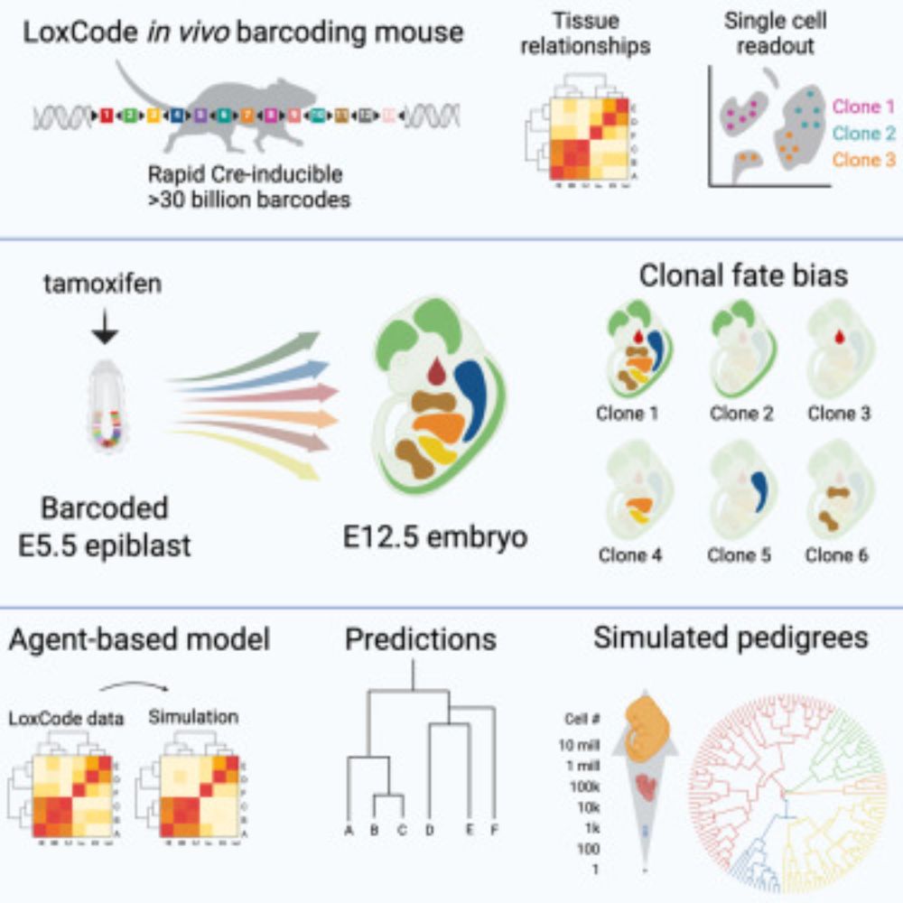
A triumph of perseverance from twitterless Tom Weber, Christine Biben and the team, our in vivo barcoding "LoxCode mouse" used to resolve epiblast fate to fetal organs is finally published in @cellpress.bsky.social and available through @jacksonlab.bsky.social www.sciencedirect.com/science/arti...
16.05.2025 04:32 — 👍 71 🔁 22 💬 8 📌 1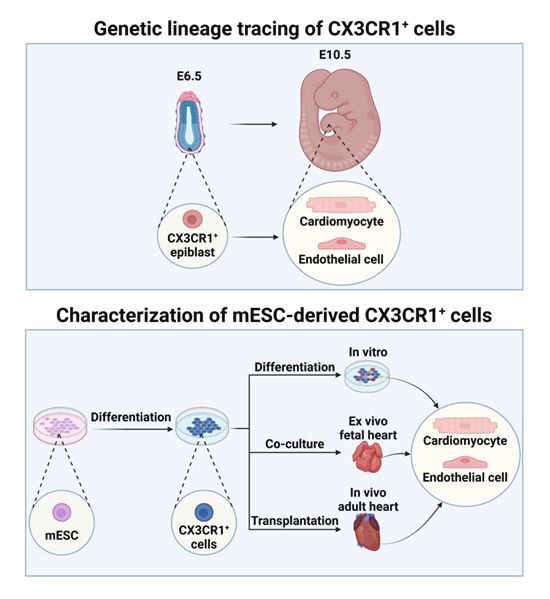
Embryonic CX3CR1+ cells constitute a multipotent epiblast-derived progenitor population that contributes not only to the formation of tissue-resident macrophages, but also of cardiomyocytes and heart endothelial cells
Young-sup Yoon and coworkers
www.embopress.org/doi/full/10....
The Company of Biologists 100 logo to the left and QR code to the right. Portrait of Helen Zenner to the left, text to the right 100 extraordinary biologists Helen Zenner Helen Zenner, Community Manager of FocalPlane and JCS Online Editor, combines her passion for microscopy, cell biology and science communication with her background in working in interdisciplinary research teams to support and connect the microscopy community. #100biologists #biologists100
This week, we are highlighting Helen Zenner, Community Manager of @focalplane.bsky.social and @jcellsci.bsky.social Online Editor, as an extraordinary biologist. #100biologists
@helenzsci.bsky.social
Sometimes authors summarize their responses to 3-6 major points raised by the reviewers in their resub cover letters or at the top of their rebuttal letters. This is so super helpful to editors, because it brings us right up to speed after not having read the paper/reviews for months. THANKS!!
09.07.2025 16:15 — 👍 189 🔁 35 💬 8 📌 4
www.biorxiv.org/content/10.1...
Boundary-guided cell alignment drives mouse epiblast maturation

3D reconstruction of a human Carnegie stage 9 embryo provides a snapshot of early body plan formation www.sciencedirect.com/science/arti...
08.07.2025 22:21 — 👍 2 🔁 1 💬 0 📌 0
Have you ever thought about inflating tissues?
Or maybe quickly deflating those inflated tissues?
New #EpithelialMechanics pre-print: doi.org/10.1101/2025...
🧵 with pressure control, multiscale buckling, controlled wrinkling
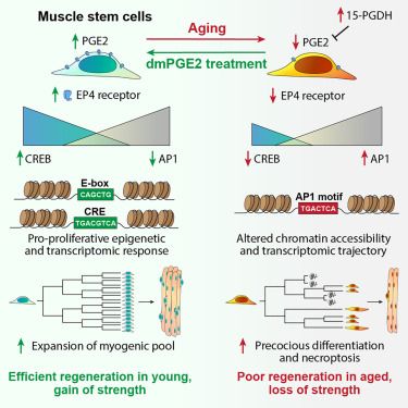
Multiomic profiling reveals that prostaglandin E2 reverses aged muscle stem cell dysfunction, leading to increased regeneration and strength
04.07.2025 14:10 — 👍 2 🔁 2 💬 0 📌 0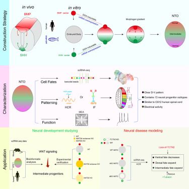
Online Now! Establishing dorsal-ventral patterning in human neural tube organoids with synthetic organizers #stemcells
14.05.2025 19:09 — 👍 1 🔁 1 💬 0 📌 0Online Now! HBO1 functions as an epigenetic barrier to hepatocyte plasticity and reprogramming during liver injury #stemcells
21.05.2025 19:09 — 👍 1 🔁 1 💬 0 📌 0
Mark your calendars for our western/evening 🌙 session seminar next week!
We’ll have a 🐁 double feature with:
Kate Cavanaugh @katecavanaugh.bsky.social and
Vera Van der Weijden @vvdw.bsky.social
🗓️ Thursday, March 20th at
⏰ 10:30 PDT (!) / 13:30 EDT (!) / 17:30 UTC / 17:30 GMT / 18:30 CET
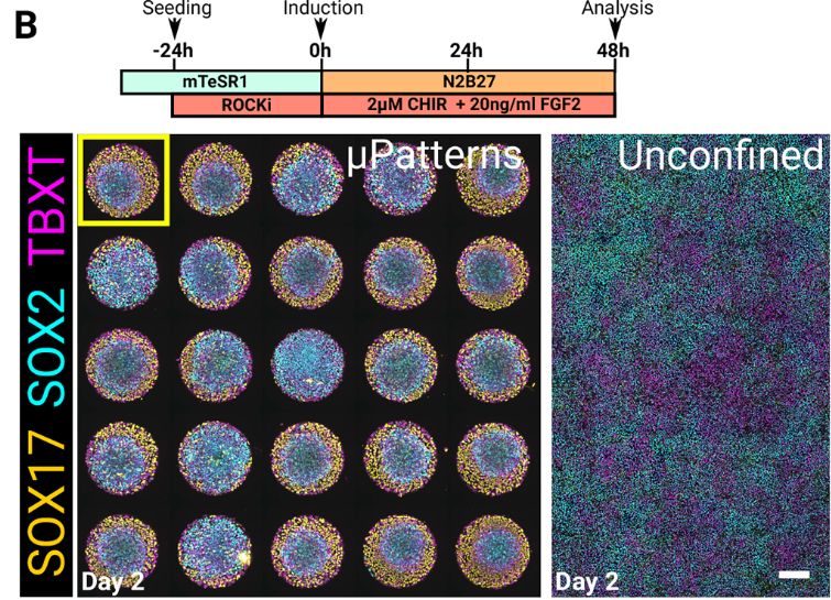
Figure 1 (B) Top: protocol used for the data shown in C-E. Bottom: confocal maximum projections showing unconstrained and micropatterned (μPatterns) cultures. Notice some variability between colonies, likely due to initial differences in seeding density across the well. The colony shown in C is indicated with a yellow outline. Scale bar: 200 µm.
In vitro modelling of anterior primitive streak patterning with human pluripotent stem cells identifies the path to notochord progenitors
Read this #OpenAccess Research Article by Miguel Robles-Garcia, Guillaume Blin and colleagues @edinburgh-uni.bsky.social:
https://buff.ly/3ZMtzc6
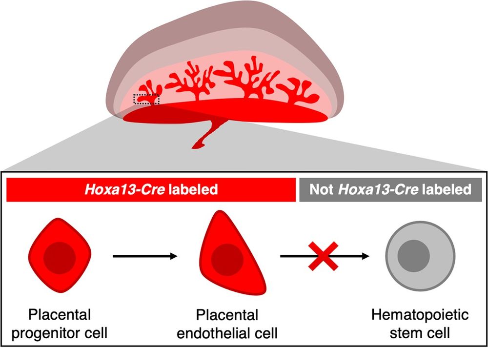
The placenta is unlikely to generate blood-forming hematopoietic stem cells (HSCs). This schematic shows how, in mouse embryos, Hoxa13-Cre lineage tracing labels virtually all endothelial cells within the mouse placenta. However, virtually no HSCs are labeled by Hoxa13-Cre. This suggests that the placenta is unlikely to form HSCs. Rather, these results suggest that HSCs are produced elsewhere in the developing embryo and subsequently migrate to the placenta, which serves as a landing pad for HSCs.
Is the placenta is a source of, or merely a niche for, blood-forming #hematopoietic #StemCells? This Primer explores a @plosbiology.org study showing that the placenta does not directly give rise to hematopoietic stem cells. 🧪 Paper: plos.io/4hl3qZF Primer: plos.io/40R1VeE
07.02.2025 10:18 — 👍 21 🔁 4 💬 0 📌 0


