Please share🙏:
Our lab opens 2 PhD positions to investigate fungi's evolution: #Barcelona
Wet lab in Biochemistry/Structural Biology of cryo-tolerant fungi
www.upf.edu/documents/d/...
Computational in Physical Chemistry of cellular processes under extrem environments
www.upf.edu/documents/d/...
26.01.2026 11:53 — 👍 16 🔁 17 💬 0 📌 3

Imaging spotlight: visualising synaptic vesicle fusion by cryo-ET - FocalPlane
Imaging spotlight: visualising synaptic vesicle fusion by cryo-ET - News
🔬🔦 Imaging spotlight on FocalPlane
@janakroll.bsky.social and colleagues highlight their research visualising synaptic vesicle fusion using cryo-ET combined with plunge freezing and optogenetics.
focalplane.biologists.com/2026/01/21/i...
22.01.2026 13:47 — 👍 12 🔁 4 💬 0 📌 0
❄️ With all the snow outside, I felt like sharing a cryo tomogram:
A neuronal synapse, captured by cryo-ET at the moment a neurotransmitter-filled vesicle fuses at the active zone.
Fusion was triggered optogenetically and arrested by plunge freezing.
More details: www.nature.com/articles/s41...
04.01.2026 08:18 — 👍 33 🔁 4 💬 0 📌 0

Der Moment, in dem eine Nervenzelle ihre Neurotransmitter in den synaptischen Spalt ausschüttet, ist extrem kurz. Berliner Forschenden @bihatcharite.bsky.social @mdc-berlin.bsky.social konnten ihn mikroskopisch einfangen.
doi.org/10.1038/s414...
Bild: © Charité/Max Delbrück Center, Jana Kroll
18.12.2025 10:04 — 👍 3 🔁 1 💬 0 📌 0
It's been a fantastic experience to publish our work in @natcomms.nature.com and I sincerely thank the reviewers of our paper for their constructive and helpful comments! (8/8)
14.12.2025 08:29 — 👍 2 🔁 0 💬 1 📌 0
I deeply thank all co-authors for their constant support over the past years: @uljakravcenko.bsky.social, @mohsensadeghi.bsky.social, Christoph Diebolder, Malgorzata Lubas, Lia Ivanov, Thiemo Sprink, Magdalena Schacherl, @mishakudryashev.bsky.social and Christian Rosenmund. (7/8)
13.12.2025 20:09 — 👍 3 🔁 0 💬 1 📌 0

Intriguingly, we observed filamentous connectors between the majority of fusing vesicles and at least one more adjacent vesicle. We suggest that these interconnectors physically couple vesicle fusion and direct vesicle resupply required for release site refilling. (6/8)
13.12.2025 20:09 — 👍 2 🔁 0 💬 1 📌 0

Our morphometric and computational analyses revealed that synaptic vesicle fusion is likely initiated via stalk formation, whereby vesicle and cell membrane form a tip-like connection. We did not find evidence for tight docking or strong membrane bending prior to fusion. (5/8)
13.12.2025 20:09 — 👍 3 🔁 0 💬 1 📌 0

In these stimulated synapses, we identified more than 50 synaptic vesicle fusion intermediates and characterized them morphometrically at nanoscale resolution and under near-native conditions. (4/8)
13.12.2025 20:09 — 👍 2 🔁 0 💬 1 📌 0

We confirmed successful optogenetic stimulation of synapses directly via cryo-confocal microscopy of the glutamate sensor iGluSnFR3. Neurotransmitters, released during exocytosis, induced an increase in fluorescence intensity that was preserved under cryo conditions. (3/8)
13.12.2025 20:09 — 👍 2 🔁 0 💬 1 📌 0

We established a timed in situ cryo-electron tomography workflow to capture the complete sequence of synaptic vesicle fusion in optogenetically stimulated mouse neurons. Using a modified plunge freezer, the "Voptobot", we stimulated neurons only 2-3 milliseconds before cryofixation. (2/8)
13.12.2025 20:09 — 👍 2 🔁 0 💬 1 📌 0
💥I am excited to present our first research paper from the lab about caveolae lipid trafficking 🎉 Led by my fantastic PhD candidate @estherocket.bsky.social
Here we looked into caveolae mediated dietary lipid uptake and how this process is mechanistically facilitated 👇 1/n
11.12.2025 07:08 — 👍 44 🔁 17 💬 6 📌 0

Postdoctoral Positions in Protein Engineering
Post a job in 3min, or find thousands of job offers like this one at jobRxiv!
We got a large Helmholtz Bioengineering grant with Oliver Daumke, Artur Yakimovich, Alina Bazarova, and Dietrich Ruess to use protein design to target cancer.
Five open postdoctoral positions in Generative AI & Protein Engineering. Share & apply!
jobrxiv.org/job/max-delb...
21.11.2025 07:08 — 👍 21 🔁 10 💬 0 📌 1
Thank you so much! 😊
01.10.2025 16:08 — 👍 0 🔁 0 💬 0 📌 0
MDC Berlin - Online Application System
Group Leader in Biomedical Engineering at the @mdc-berlin.bsky.social in Berlin.
It's a broad call for whoever is doing exciting Bioengineering, please apply or share. We have a great institute with amazing colleagues and core facilities,highly recommend
application.mdc-berlin.de/site/index.p...
01.10.2025 15:39 — 👍 7 🔁 6 💬 1 📌 0
Postdoc program at the MDC in Berlin, @mdc-berlin.bsky.social : take a look 👇👇
For this call, collaborative projects between two groups will be funded. Take a look at the call and the groups and drop me an email if interested. Share with colleagues too!
www.mdc-berlin.de/postdocs#t-g...
04.08.2025 10:23 — 👍 11 🔁 8 💬 0 📌 0
scientific image
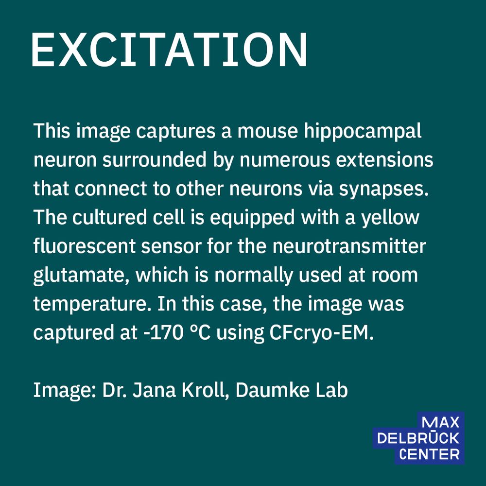
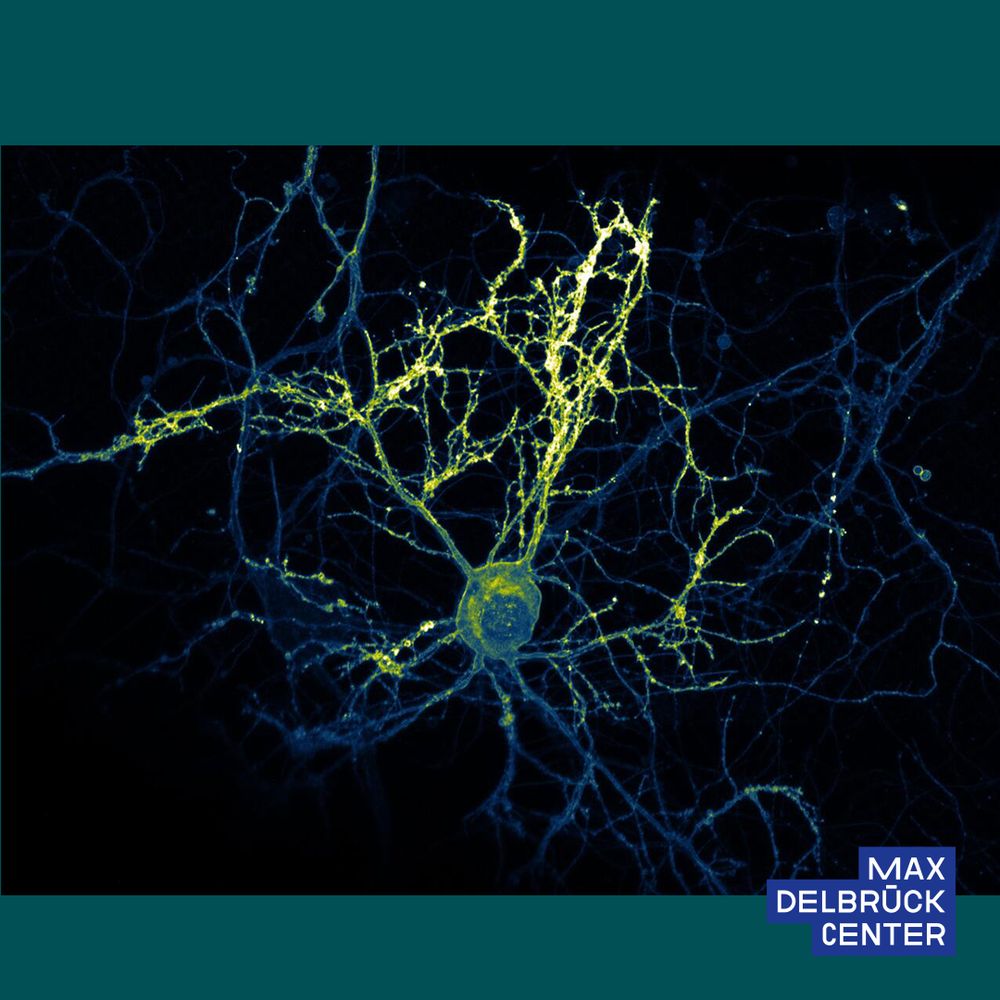
Excitation
This image captures a mouse hippocampal neuron surrounded by numerous extensions that connect to other neurons via synapses. The cultured cell is equipped with a yellow fluorescent sensor for the neurotransmitter glutamate, which is normally used at room temperature. In this case, the image was captured at -170 °C using CFcryo-EM (Core Facility for Cryo–Electron Microscopy).
Image: Dr. Jana Kroll, Daumke Lab
scientific image
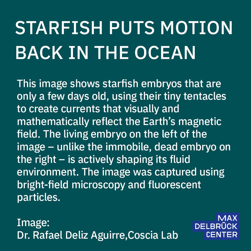
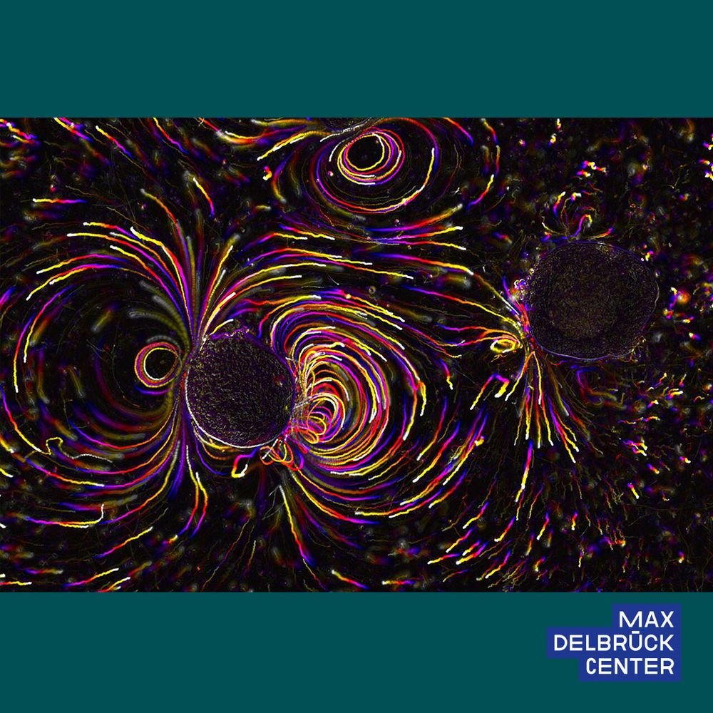
Starfish Puts Motion Back in the Ocean
This image shows starfish embryos that are only a few days old, using their tiny tentacles to create currents that visually and mathematically reflect the Earth’s magnetic field. The living embryo on the left of the image – unlike the immobile, dead embryo on the right – is actively shaping its fluid environment. The image was captured using bright-field microscopy and fluorescent particles.
Image: Dr. Rafael Deliz Aguirre, Coscia Lab
🏆 Images from #mdcBerlin received top honors in the @helmholtzimaging.bsky.social “Best Scientific Image Contest”!
Jury Award for @janakroll.bsky.social, Daumke Lab
Public Choice Award for Dr. Rafael Deliz Aguirre, @coscialab.bsky.social
Full details: www.mdc-berlin.de/news/news/tw...
17.07.2025 13:38 — 👍 18 🔁 5 💬 0 📌 0
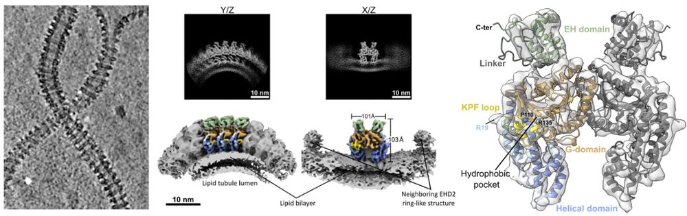
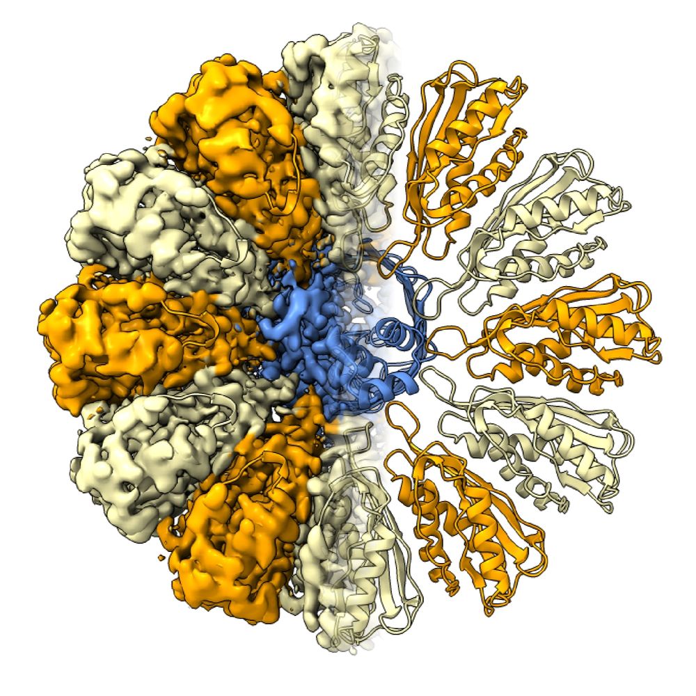
We have a preprint for you
EHD2 forms rings on caveolae necks, in contrast to most EHDs forming helices. We determined its structure on membranes and show that N-term acts as a spacer
By Elena, Vasya @vasiliimikirtumov.bsky.social, Jeff &Oli Daumke www.biorxiv.org/content/10.1...
08.06.2025 11:21 — 👍 107 🔁 34 💬 4 📌 0


What an inspiring morning at the CatCat Symposium! Thank you so much for inviting me @gallegolab.bsky.social and @felixmendu.bsky.social, it's been a pleasure visiting you in Barcelona 😊
27.05.2025 18:03 — 👍 11 🔁 3 💬 2 📌 0
In the left image, I see a queen with a crown🫅 But I guess that's not what you meant? 😅
13.05.2025 17:46 — 👍 2 🔁 0 💬 0 📌 0
So many amazing images! 🤩 🤩🤩
Please make sure to vote, mine is also in the list 😎
12.05.2025 18:11 — 👍 1 🔁 0 💬 0 📌 0

PhD /Postdoc in In Situ Structural Biology by Cryo-ET
Post a job in 3min, or find thousands of job offers like this one at jobRxiv!
We are looking for a PhD student/Postdoc to develop methods of cryo-ET and/or study the structure and function of membrane proteins. Great team at the MDC in Berlin.
It's a broad call, please apply and share!
jobrxiv.org/job/max-delb...
28.03.2025 08:42 — 👍 20 🔁 13 💬 0 📌 0
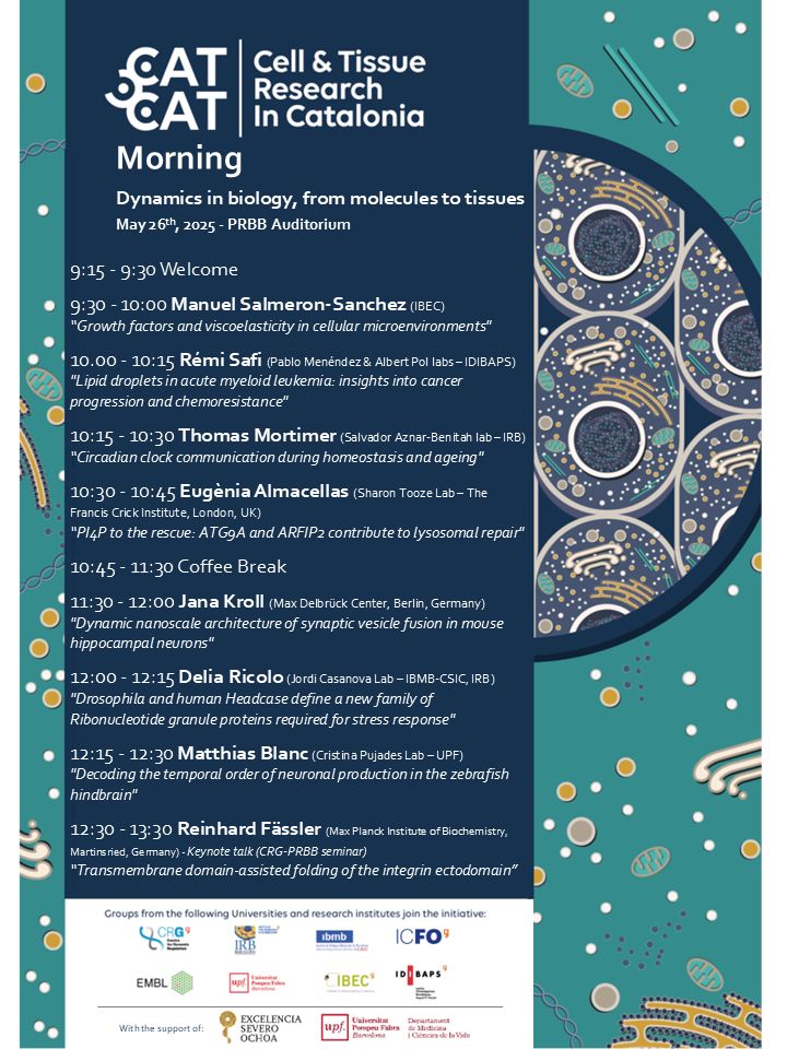
Program of the 9th CATCAT morning on "Dynamics in biology, from molecules to tissues", May 26th 2025
Together with @gallegolab.bsky.social we are organizing the next CATCAT morning on "Dynamics in biology, from molecules to tissues", with an amazing lineup of local and international speakers. If in Barcelona on May 26th, don't miss it, it's free! www.catcat-celltissuebiology.cat/event/9th-ca...
14.03.2025 09:57 — 👍 11 🔁 11 💬 2 📌 0

Cryo-confocal microscopy of a neuron expressing the glutamate sensor iGluSnFR 🥶 The neuron was optogenetically stimulated and subsequently plunge-frozen to preserve the high fluorescence intensity indicating neurotransmitter release. #FluorescenceFriday
21.02.2025 08:05 — 👍 65 🔁 8 💬 0 📌 0
Thank you!
16.02.2025 21:09 — 👍 1 🔁 0 💬 0 📌 0
The video shows more than 50 snapshots of vesicle fusion events in synapses, sorted by fusion progression 😎
15.02.2025 20:16 — 👍 0 🔁 0 💬 0 📌 0
Thank you! 😊
14.02.2025 08:58 — 👍 0 🔁 0 💬 0 📌 0
Joint PhD Student at the Max-Planck-Institutes for Biophysics and -chemistry
#TeamTomo #CryoEM #Microscopy #Microfluidics #Vitrification
Postdoc at the University of Bristol, studying the endosomal system using biochemistry and electron microscopy 🔬
Previously DPhil student at the University of Oxford working on viral genome replication 🦠
https://orcid.org/0000-0002-9650-382X
Marie Curie Postdoc Fellow at Utrecht University 🇳🇱 Protein-Biochemist diving into cryo-ET ❄️
Postdoctoral Scholar in the Barad Lab at Oregon Health and Science University, where I study cytoskeletal remodeling in response to intracellular infection using Cryo-electron tomography. Views are my own.
Former PhD student in the Carter lab (MRC LMB), now postdoc in the Beck lab (MPI of Biophysics).
PhD candidate @institutpasteur | Piplettes_mag✍🏻
Structural biologist studying proteins that move things from A to B. University of Queensland, Institute for Molecular Bioscience. Centre for Cell Biology of Chronic Disease.
He/Him.
https://imb.uq.edu.au/research-groups/collins
Shooting electrons, ions and photons (mainly) at plants to study cell-cell communication @mpibiochem.bsky.social & @hhu.de
Postdoc @JohnBriggsGroup @MaxPlanck Institute of Biochemistry in Martinsried
| PhD from PlevkaLab | #StructuralBiology of #Viruses #HIV | #CryoEM | #Memes
Twitter/X: @F4ustus
Staff #scientist in the Landau group (https://landau-lab.de) at CSSB/DESY, structural biologist studying functional #amyloids using #cryoEM.
Climbing in my spare time.
🇸🇪 in 🇩🇪
PhD student in structural biology
Computational Cryo-EM/ET at Genentech
Love people, the outdoors and building stuff
Complexity is the enemy
All opinions my own
BIOspektrum - Das Magazin für Biowissenschaften * Neue Themen aus der Wissenschaft * Nachrichten aus der Scientific Community
http://www.biospektrum.de
🧫 Join 700+ researchers getting weekly lab hacks with our newsletter (it’s free) 👉 wildtypeone.substack.com/about
"Synthetic Membrane Systems" group
Biochemistry, University of Düsseldorf
Membrane proteins, Good spirit and some Anarchy
https://isb.med.upenn.edu/
Nature Communications is an open access journal publishing high-quality research in all areas of the biological, physical, chemical, clinical, social, and Earth sciences.
www.nature.com/ncomms/
👩🔬 The Institut Pasteur is a leading global biomedical research institute.
🔬 Explore with us the frontiers of biomedical research
🧪 Follow us to uncover groundbreaking discoveries
🌍 Join a community passionate about progress and open science
Deutsche Gesellschaft für Zellbiologie | German Society of Cell Biology https://www.zellbiologie.de
PhD student at Karolinska Institutet and Scilifelab | Cell physics
https://www.csi-nano.org/