Last days to apply this position in our group! Do join us in this great Institute @crg.eu, with great colleagues, and help shaping the science and culture of our lab!
09.02.2026 14:24 — 👍 9 🔁 10 💬 0 📌 0
Thank you A. Perez-Ramos and P. Bovolenta for this great perspective on our article about Optic Cup morphogenesis @dev-journal.bsky.social !
26.11.2025 13:22 — 👍 4 🔁 2 💬 0 📌 1
Congrats ☺️☺️
04.09.2025 14:31 — 👍 1 🔁 0 💬 1 📌 0
Fantastic interaction with @carldmodes.bsky.social
and @liormoneta.bsky.social , who did all the modelling. It was great to work in such an interchangeable setting between biology and physics, to lear about #ShapeProgramming and shed a little more light on how organs gain their 3D shape.
01.09.2025 21:22 — 👍 2 🔁 1 💬 0 📌 0
This work also had the great contributions of A. Szalapak,
@louise-dagher.bsky.social and
@the-chaotician.bsky.social . Special thanks also to
@lcferme.bsky.social for the stardist model! And of course to Caren and all the @nordenlab.bsky.social
01.09.2025 21:22 — 👍 0 🔁 0 💬 1 📌 0
Finally, we disrupted apical patterned behaviors experimentally. The optic cup completely failed to start the invagination! (side note, the phenotype was so striking that I thought the microscope had crashed while imaging)
01.09.2025 21:22 — 👍 1 🔁 0 💬 1 📌 0

Our model became even cooler: we converted tissue segmentations into networks. Like this we could test the strain patterns in the REAL geometry of the tissue
01.09.2025 21:22 — 👍 1 🔁 0 💬 1 📌 0

But can the shape transitions at the apical surface be sufficient to drive basal surface invagination? Updating our model with basal surfaces: apical strain patterns alone are enough to initiate invagination—even if basal surfaces are passive
01.09.2025 21:22 — 👍 0 🔁 0 💬 1 📌 0

First, we needed to understand how are the apical surfaces curving. We went back and forth between experiments and modelling (inspired by shape programming). We found each apical surface displays different cell behaviors, generating its own in-plane strain patterns,
01.09.2025 21:22 — 👍 0 🔁 0 💬 1 📌 0
But the surprise came from the apical surfaces. They undergo shape transitions before invagination starts. Could they be driving the process?
01.09.2025 21:22 — 👍 0 🔁 0 💬 1 📌 0
Separating the basal surface of the two epithelia (NE and RPE), we saw that they change their shape at the same time!
01.09.2025 21:22 — 👍 0 🔁 0 💬 1 📌 0
We followed 3D shape changes of the forming optic cup, by tracking the basal surface
01.09.2025 21:22 — 👍 0 🔁 0 💬 1 📌 0
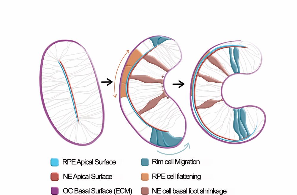
Many things happen at the same time: 2 epithelial layers differentiate, cells migrate, other cells flatten and others constrict their basal surface, and the lens grows in. We went through (a LOT of) negative results to try to understand which processes were triggering the onset of the invagination
01.09.2025 21:22 — 👍 0 🔁 0 💬 1 📌 0
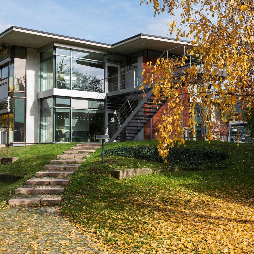
We used the Optic Cup formation as a system. The shape transition from a flat vesicle to an hemispheric eye precursor is just mesmerizing
01.09.2025 21:22 — 👍 0 🔁 0 💬 1 📌 0
We departed from a bigger question: how 3D shape emerges in complex tissues, from the interaction of multiple cell behaviors and mechanical inputs?
01.09.2025 21:22 — 👍 0 🔁 0 💬 1 📌 0
Briefly, we found that OC invagination onset relies on active, collective cell behaviors, that create patterned strains at the apical surfaces. In addition, OC morphogenesis involves the cooperative reinforcement of two independent active patterns. More details bellow 👇
01.09.2025 21:22 — 👍 0 🔁 0 💬 1 📌 0
Training 12 doctoral candidates in a new field of comparative integrative cell biology in animals. Funded by the European Union. GA: 101119891
[bridged from https://biologists.social/@ZooCELL on the fediverse by https://fed.brid.gy/ ]
Dissecting psychiatric disorders using frogs (she/her)
willseylab.com
Co-Creating Ireland's Public Involvement in Open Research Roadmap
ENGAGED is building a national roadmap to shape public involvement in open research in Ireland. We believe that research can and does play an important role in tackling societal challenges.
Monthly online seminar series at the intersection of metabolism, developmental biology & organismal physiology. Every 2nd Thursday of the month at 16:00 CET.
Subscribe at: https://forms.gle/Y8QzucogKKrZ6zYv7
MPI-CBG, Paul Langerhans Institute (PLID) -Developmental and stem cell biologist- Physics of Life
Microbiologist 🇵🇹 @cnrs.bsky.social, @ENS-Lyon, @igfl.bsky.social, @LeulierLab #Probiotics #Growth #Prophages #Stress #Drosophila
Physicist having a go at biology, EMBO postdoctoral fellow @UCL in the Mao group, @EMBL alumna
Assistant Prof @univmiami.bsky.social. Cellular and Developmental Biology / Evo-Devo / Animal Multicellularity / Cell adhesion. Lover of weird invertebrates. Learn more: clarkelab.com
The wanderer. The clown. The chairman.
President and CEO, @alleninstitute.org
Opinions are my own.
Group leader SNSF Ambizione at EPFL, Lausanne. Formerly, Postdoc Roux lab, Geneva; PhD Piel lab, Paris. #cytoplasts #actomyosin #cellbio #microscopy #membranes #biophysics #extracellular_vesicles #sciart
Lab website: http://celldynamicslab.com
Account of the Max Planck Institute for the Physics of Complex Systems in Dresden; tweets by Pablo Pérez, Uta Gneisse, and Pierre Haas @lepuslapis.bsky.social.
Professional nerd (science journalist). USian in Austria, language geek, and collector of fine yellow zigzagged sweaters and etymology fun facts. Get my newsletter about big questions at the frontiers of science: www.reviewertoo.com 👽🌀🦋
MorphoNet is an interactive viewer dedicated to 3D and 3D+t datasets made with intensity images and/or segmented data.
Non-profit organisation - Champalimaud Centre for the Unknown
Neuroscience & Cancer Research
Science Communication & Outreach
https://www.fchampalimaud.org/champalimaud-research
PhD Student@University of Sussex w/ Tom Baden
MSc Neuroscience@University of Tübingen w/ Thomas Euler
Interested in visual neuroscience, neural design, evolution.
Optical physicist. Microscopy and biophysics researcher. Currently postdoc @HenriquesLab 🔬
I like bicycles 🚲
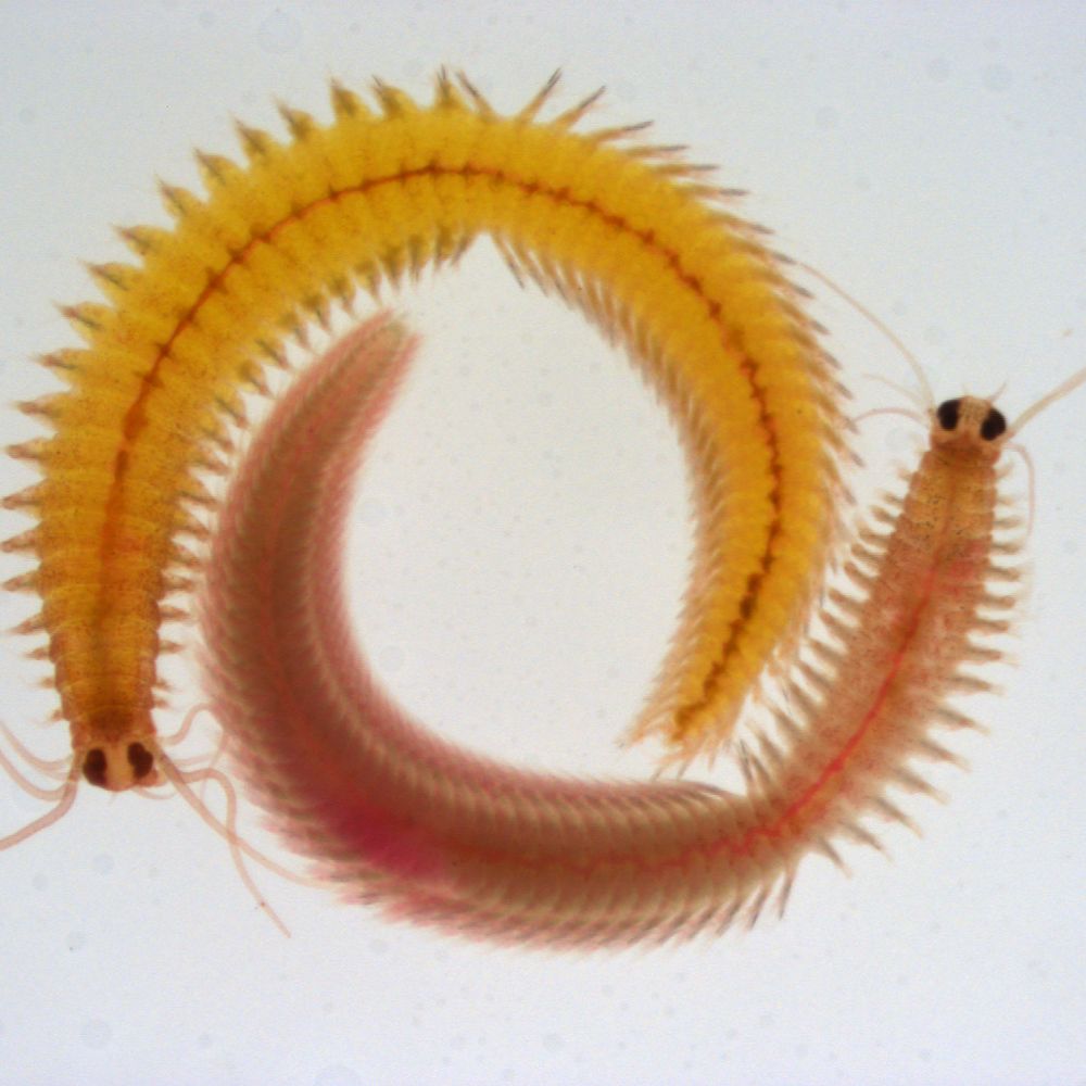
Shared account Tessmar & Raible labs, University of Vienna @univie.ac.at and AWI. Marine clocks and rhythms, moon- and sun light, stem cells, hormones, regeneration and biomaterials.
Post-Doc @katjaroeper.bsky.social lab @pdncambridge.bsky.social
Interested in kidney organoids and lumen formation; Former @honigmann_lab,
@biotec-tud.bsky.social, @cmcb-tud.bsky.social,@mpicbg.bsky.social
and @boulantlab
Cutting-edge research, news, commentary, and visuals from the Science family of journals. https://www.science.org