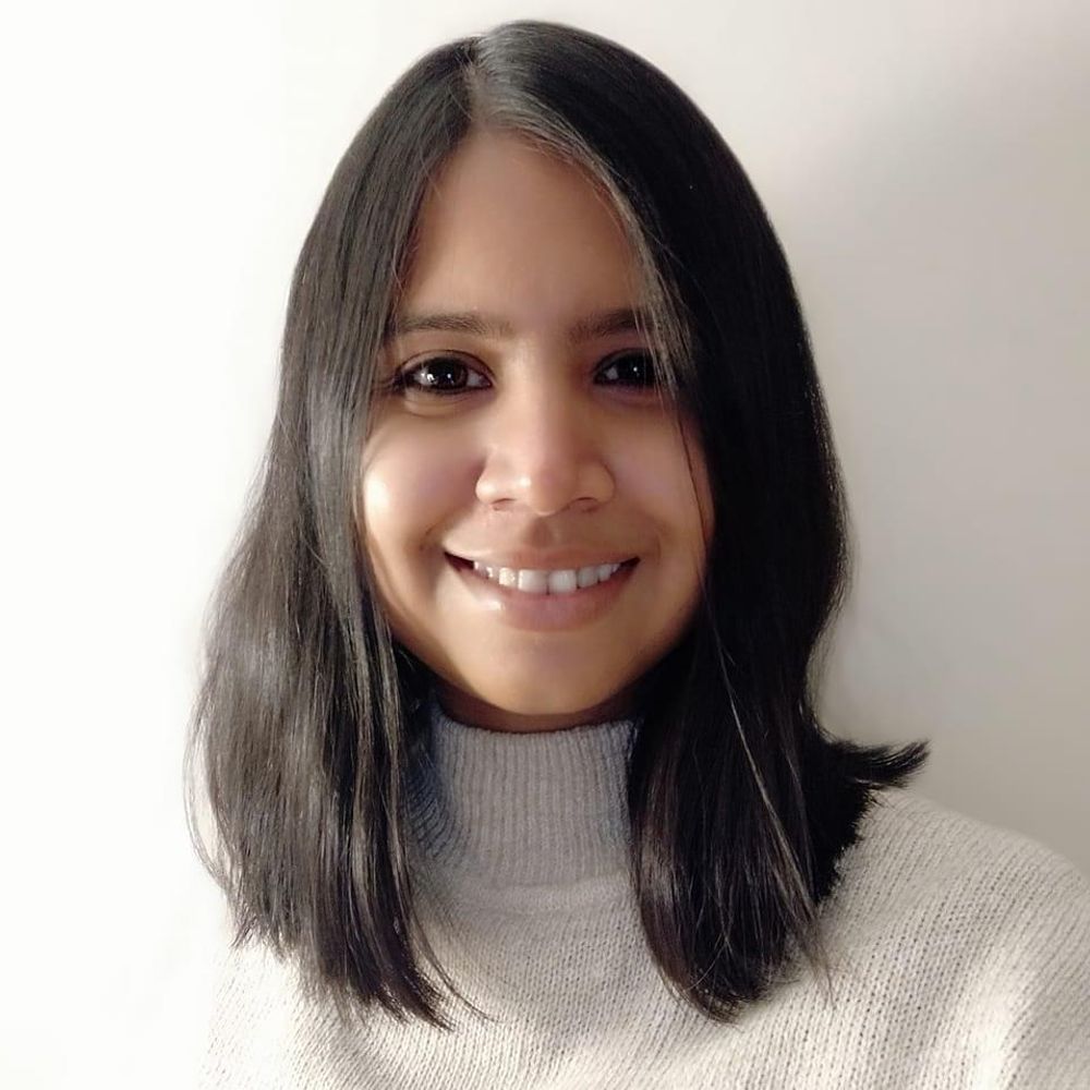
AJs maintain tissue integrity by maturing in response to actomyosin generated tension at cell–cell contacts. (A) AJs are formed in the apical region of polarised epithelial tissues. Subsequent figures show a top-down view of an x-y plane at the apical region. (B) Schematic defining the key components involved in cell–cell adhesion, as discussed in this Review. Cadherin–catenin complexes (CCCs) hold together the actin cytoskeletons of adjacent cells at regions of cell–cell contact. Clustering of CCCs forms AJs. (C) Pulling forces exerted on CCCs changes their bio-mechanical properties. The E-cadherin trans interaction changes from an X- (left) to a strand-swap (right) conformation and α-catenin recruits vinculin, which strengthens the binding of α-catenin to F-actin. Florette indicate catch bonds, which increase in lifetime with applied force (E-cadherin X dimer, α-catenin tail domain–actin and vinculin tail domain–actin). Created in BioRender by Winn, L., 2025. https://BioRender.com/l1sc933. This figure was sublicensed under CC-BY 4.0 terms.
In their Review, John James, Darius Köster and colleagues consider E-cadherin-mediated cell-cell adhesion, focusing on recent advances in our understanding how actin cortex dynamics affect adherens junction stability.
journals.biologists.com/jcs/article/...
09.12.2025 10:06 — 👍 14 🔁 5 💬 0 📌 0
LinkedIn
This link will take you to a page that’s not on LinkedIn
Our Lab @ NCBS, Bangalore is hiring PhD students!
If you’re excited about cell & developmental biology, tissue mechanics, and imaging — come join us! We use C. elegans to uncover how forces shape organs 🧫🔬
CSIR/DBT/ICMR fellowship holders encouraged to apply!Apply 👉 lnkd.in/gr9EUHnp
08.12.2025 10:55 — 👍 8 🔁 7 💬 0 📌 0
#aECM at #CellBio2025 On Sunday, the Sundaram lab will have 2 posters on #Celegans aECM - boards 223 and 225
06.12.2025 20:07 — 👍 10 🔁 3 💬 1 📌 0

Epithelial Mechanics Fan Club - FocalPlane
Epithelial Mechanics Fan Club - News
Meet the Epithelial Mechanics Fan Club @epimechfc.bsky.social
Nimesh Chahare @onenimesa.bsky.social and Julia Eckert @juliaeckert.bsky.social introduce us to the Epithelial Mechanics Fan Club.
Learn how you can get involved with this community: focalplane.biologists.com/2025/12/04/e...
05.12.2025 13:35 — 👍 13 🔁 8 💬 0 📌 2

The Larry Sandler Award recognizes excellent recent graduates who have completed a PhD in Drosophila research. The awardee will receive a fantastic opportunity to present the Larry Sandler Memorial Lecture at the 67th Annual Drosophila Research Conference in Chicago, IL!
The deadline to nominate is December 15, 2025.
Nominate Here
Eligibility
Any student completing a PhD in an area of Drosophila research between July 2024 and December 2025 is eligible. Students may be nominated by their thesis advisor, department chair, or supervisory committee member.
Nominations
Nominations may be submitted by the student’s thesis advisor, department chair, or supervisory committee member, and should include the following:
Nominee's CV
Nominee's thesis abstract of 1–2 pages
A nomination letter directly answering the following questions:
What are the main discoveries in the nominee's thesis?
What are the intellectual and experimental contributions of the nominee to the project?
How does the thesis work advance what was previously known in the field and to previous work from your lab?
Drosophila colleagues--Please submit nominations for the Larry Sandler Award. which recognizes excellent recent graduates who have completed a PhD in Drosophila research. Details are below and at this link:
genetics-gsa.org/drosophila-2...
04.12.2025 16:41 — 👍 9 🔁 16 💬 0 📌 0
🚨 New preprint!
We built a single-cell atlas of 14 multilayered epithelia and revealed a conserved transcriptomic program guiding tissue architecture and fate composition. Our work brings decades of tissue-specific studies together into a unified evo-devo framework.
www.biorxiv.org/content/10.1...
17.11.2025 12:32 — 👍 40 🔁 12 💬 1 📌 0

New paper from the @rouxlab.bsky.social on Nature Communications! We study how membrane tension is spatially organized in cells. Using the mechanosensitive probe Flipper-TR to visualize tension across the plasma membrane of adherent cells and to dissect the conditions needed for a gradient to happen
27.11.2025 14:14 — 👍 83 🔁 32 💬 6 📌 2

The historc city center of Münster, famous for the Peace of Westphalia!
2/2 If you’re into #cellbio, cellular interfaces, #mechanobiology, #membranes, anything related, come to Münster, Germany, May 27–29!
www.uni-muenster.de/SFB1348/en/i...
Please spread the word. It’s a fantastic (and FREE!) meeting. And while you’re here, explore Münster… it’s worth it.
#Science
28.11.2025 08:33 — 👍 17 🔁 11 💬 0 📌 0
Ditipriya Mallick, Siddhartha Sankar Jana and colleagues discover that cellular elasticity drives mechano-adaptation against fluid shear stress.
journals.biologists.com/jcs/article/...
27.11.2025 08:04 — 👍 7 🔁 3 💬 2 📌 0

This artwork was inspired by the ancient Chinese legend of "The Cowherd and the Weaver Girl," where two star-crossed lovers are reunited once a year by a bridge of magpies. Here, the Cowherd and Weaver Girl represent two non-interacting proteins (e.g., actin and YAP1), separated by the Milky Way—symbolizing the complex cellular environment. The magpie bridge, formed by STUPPIT components with Pup(E) as the body and Split-TurboID fragments as glowing wings, enables their connection. Soaring lanterns released from the bridge represent biotin-labeled proximal proteins (e.g., AMOT), illuminating the functional interactome between the once-distant proteins. Xie et al. demonstrate how the STUPPIT system acts as a molecular "magpie bridge," enabling proximity labeling and revealing functional interactomes between proteins that do not directly bind—just as the magpie bridge reunites the star-crossed lovers. Artwork by graduate student Xin Li.
Despite advances in #ProximityLabeling tools, we can't capture intermediary proteins that bridge two associated non-interacting proteins. STUPPIT is a novel method to label intermediary proteins between two non-interacting partners in signaling pathways @plosbiology.org 🧪 plos.io/4ab6YwQ
27.11.2025 08:55 — 👍 3 🔁 2 💬 0 📌 0

So called microtubule organizing centers (MTOCs) are not "organizing centers". They don't control their position (network center), nor MT orientation, length and shape (network shape). The actual organizers are MTs, molecular motors and regulators of MT +ends. MTOCs nucleate MTs.
Let's rename MTOCs.
27.11.2025 08:47 — 👍 67 🔁 16 💬 10 📌 0
Gorgeous website, Maik!
27.11.2025 14:31 — 👍 1 🔁 0 💬 1 📌 0
Prospective PhD students if you are looking to work on some exciting Developmental Biology questions combining some state-of-the-art microscopy, please consider joining the talented @maikbischoff.bsky.social
27.11.2025 14:30 — 👍 8 🔁 3 💬 1 📌 0

The retina’s rhythm
Calcium waves facilitate the emergence of sight
Check out my Perspective article “The Retina’s Rhythm” which accompanies an exciting paper by Claude Desplan in Science. I highlight his discovery that waves of calcium are required to finalize the honeycomb-like structure of the Drosophila retina.
www.science.org/doi/epdf/10....
21.11.2025 14:00 — 👍 20 🔁 6 💬 0 📌 0
Good job and best wishes, Shefali! 🎉
26.11.2025 12:57 — 👍 1 🔁 0 💬 1 📌 0
Super proud to be part of SFB1348 — and excited to share that I now officially have my own lab in Münster! 🎉🪰
We’ll study #cellbio & #morphogenesis, focusing on organ sculpting and #chirality in #Drosophila.
I’ll soon hire my first #PhD student — feel free to reach out!
#juniorPI #Science #devbio
25.11.2025 15:56 — 👍 103 🔁 21 💬 24 📌 1
Onwards and upwards! Congrats Maik 🎉
26.11.2025 12:51 — 👍 1 🔁 0 💬 1 📌 0
Well deserved, Ben! 🎉
21.11.2025 12:56 — 👍 2 🔁 1 💬 0 📌 0
Attempt number seven at uploading this video of intermediate filaments in an enormous COS7 cell. I have a feeling the BlueSky compression will not do it any favors.
21.11.2025 03:54 — 👍 942 🔁 82 💬 98 📌 12
I am very excited to be organizing a session for postdocs (with focus on those in the job market). Be on the lookout for info and registration! 🐟🧪
19.11.2025 19:12 — 👍 45 🔁 13 💬 3 📌 0

SAVE THE DATE! Stoked to organize the 2026 Santa Cruz Developmental Biology Meeting with @rashmi-priya.bsky.social, @lowelab.bsky.social, and Shelbi Russell. Come learn about Biomedicine, Biomechanics, and the Biosphere, August 24-28, 2026. Registration dates, etc., coming soon! Please RT
19.11.2025 20:23 — 👍 115 🔁 74 💬 1 📌 3
#aECM Cub starts next week Nov 18 with talks on patterning the Drosophila lens, mouse tectorial membrane, and C.elegans cuticle furrows. You can still sign up for access using the link below.
14.11.2025 11:56 — 👍 7 🔁 4 💬 1 📌 0

How do four-eyed fish see above & underwater? 🌤️🌊 Our new preprint reveals how Anableps rewired its retina for dual vision- evolution at work 👁️👁️ Kudos to @perezlouise.bsky.social @josanesousa.bsky.social @keylapruett.bsky.social + team!
🔗 tinyurl.com/3a8r9xy5
04.11.2025 15:56 — 👍 39 🔁 19 💬 1 📌 0

Volume 152 Issue 20 | Development | The Company of Biologists
Development that lasts a life time. Check out the 2025 special issue of @dev-journal.bsky.social on Lifelong Development: the Maintenance, Regeneration and Plasticity of Tissues
journals.biologists.com/dev/issue/15...
05.11.2025 08:54 — 👍 28 🔁 15 💬 0 📌 0
Neuroscientist and glial aficionado at NYU Grossman School of Medicine/NYU Langone Health in NYC. Posts in my individual/personal capacity.
My lab is full of awesome people doing amazing stuff - check them out: www.liddelowlab.com
he/him
Biologist; Umass Chan Medical School
Group leader Ambizione at EPFL, Lausanne. Formerly, Postdoc Roux lab, Geneva; PhD Piel lab, Paris. #cytoplasts #actomyosin #cellbio #microscopy #membranes #biophysics #extracellular_vesicles #sciart
Lab website: http://celldynamicslab.com
Dev cell syst biol, cardiopharyngeal, coll migr, ecodevo. Dir Sars Centre, U Bergen. NIH DEV1 rev. Prof NYU Bio. Postdoc UC Berkeley. PhD CNRS/Paris XI. Agrégé. Archicube. French & American.
https://www.uib.no/en/michaelsarscentre/141681/christiaen-group
Developmental biologist | Professor at Indiana University Department of Biology | eye development, pattern formation, gene regulation
Assistant Professor at @DukeBiology. #NewPI #EvoDevo, gene regulation, adaptive traits, #Wnt signaling 🦋. 🧬🔬🧪. She/her/Ella
Collaborative Research Center investigating the formation and function of dynamic cellular interfaces at the University of Münster, Germany. Funded by the German Research Foundation (DFG).
https://www.uni-muenster.de/SFB1348/en/index.html
Automatically posts newly uploaded #Drosophila papers from Pubmed every hour.
Admin: @tkmkmym.bsky.social
Monthly online seminar series at the intersection of metabolism, developmental biology & organismal physiology. Every 2nd Thursday of the month at 16:00 CET.
Subscribe at: https://forms.gle/Y8QzucogKKrZ6zYv7
Asst Professor at LSU. Four-eyed fish evodevo. Education and Professional Training Officer- PANAM Evodevo Society.
A project highlighting the stories and careers of women in neuroscience 👩🏾🔬👩🔬👩🏿🔬👩🏻🔬
Podcast here http://linktr.ee/storiesofwin
Neuroimmunology & Bioinformatics 🦠 | Schmidt Science & Weill Neurohub Postdoctoral Fellow | SENse Lab @berkeleymcb.bsky.social | PhD in the deadliest animal 🦟 | Co-founder & Board Secretary @cientificolatino.com 💗 | she/her 🏳️🌈
Comparative developmental biology, regeneration, non-conventional model organisms, live imaging; see www.averof-lab.org
RNA, Cell Biology, Zebrafish, Development Lovers
Professor at University of California, Davis. Studying cadherins, desmosomes, biophysics of cell adhesion. Also developing new proximity labeling technologies.
https://sivasankarlab.ucdavis.edu/
Synthetic developmental biologist at PoL TU Dresden. Cross-species comparison and manipulation of the ORGANOID ZOO.
https://physics-of-life.tu-dresden.de/team/pol-groups/ebisuya
Developmental biology, University of Cambridge
https://www.gurdon.cam.ac.uk/people/emma-rawlins/
Physicist having a go at biology, EMBO postdoctoral fellow @UCL in the Mao group, @EMBL alumna
Principles of Tissue Morphogenesis @Duke
https://www.munjallab.com/