Super excited we made the cover of @natcardiovascres.nature.com! Work represents ADAR1 RNA editing within the vascular wall
15.10.2025 13:38 — 👍 2 🔁 0 💬 0 📌 0
Excited for a major milestone in our efforts to map enhancers and interpret variants in the human genome:
The E2G Portal! e2g.stanford.edu
This collates our predictions of enhancer-gene regulatory interactions across >1,600 cell types and tissues.
Uses cases 👇
1/
18.09.2025 16:14 — 👍 84 🔁 36 💬 2 📌 1
Hey thanks! Hope you’re doing well!
17.09.2025 01:22 — 👍 0 🔁 0 💬 0 📌 0

There's a lot here and a lot more in the paper. But I get excited thinking about the potential. From rare to complex disease to novel mechanisms with real potential for a precision guided approach to therapy. A lot to do! @stanfordmedicine.bsky.social @stanforddeptmed.bsky.social
16.09.2025 12:47 — 👍 2 🔁 1 💬 0 📌 0

To then connect this back to humans, entirely grateful for collaborators @clintomics.bsky.social & Sander van der Laan where we investigated ISG activation and SMC modulation and plaque phenotype in the Athero-Express cohort, showing distinct relationships between ISG induction and calcification
16.09.2025 12:47 — 👍 0 🔁 0 💬 1 📌 0

Importantly, we define the cellular trajectory of MDA5 activation leading to vascular calcification and disease progression, an effect that can be entirely inhibited with simply haploinsufficiency of MDA5.
16.09.2025 12:47 — 👍 0 🔁 0 💬 1 📌 0

This MDA5 activation leads to increased plaque size due to increased SMC migration into the plaque with markedly increased vascular calcification.
16.09.2025 12:47 — 👍 0 🔁 0 💬 1 📌 0

But importantly, we show that with SMC ADAR1 haploinsufficiency, atherosclerosis studies reveal that MDA5 activation occurs in a cell type and context specific mechanism. MDA5 activation drives a distinct SMC cell state change.
16.09.2025 12:47 — 👍 0 🔁 0 💬 1 📌 0

In atherosclerosis - we show that SMCs appear to be enriched for these immunogenic RNA, and that as SMC undergo phenotypic modulation in both human and mouse there is significant activation of ISG genes, potentially suggestive of MDA5 activation
16.09.2025 12:47 — 👍 0 🔁 0 💬 1 📌 0

With homozygous deletion of ADAR1 in SMC, there is a loss of vascular integrity. Further single cell RNA sequencing reveals distinct ISG activation and cellular infiltration with critical receptor ligand interaction
16.09.2025 12:47 — 👍 1 🔁 0 💬 1 📌 0

Our work here gets at this mechanism.
We reveal a fundamental observation, that vascular SMC have a unique requirement for ADAR1 editing to prevent MDA5 activation.
SMC deletion of ADAR1 leads to severe phenotype within days and is entirely blocked with deletion of MDA5
16.09.2025 12:47 — 👍 0 🔁 0 💬 1 📌 0
In addition, rare LOF variants in MDA5 (IFIH1) have been found to be protective against CAD as well as other inflammatory disorders. This provides quite strong human genetic evidence to support ADAR1-dsRNA-MDA5 axis in CAD, but through what mechanism?
16.09.2025 12:47 — 👍 0 🔁 0 💬 1 📌 0

The big finding in 2022 by my colleagues Qin Li (now @upenn.edu and Billy Li @stanforduniversity.bsky.social)
was that beyond rare disease, common variants appear to regulate RNA editing (edQTLs), and these edQTLs predict numerous common inflammatory disorders, including CAD! t.co/t1i47lPlGG
16.09.2025 12:47 — 👍 0 🔁 0 💬 1 📌 0

Amazingly, mice that are deficient in ADAR1 are embryonic lethal, but dual knock out of ADAR1 and MDA5 essentially rescues the phenotype. In this case, the role of ADAR1 seems to be nearly entirely based on preventing MDA5 activation, less so the actual edit of the transcript
16.09.2025 12:47 — 👍 0 🔁 0 💬 1 📌 0

In rare disease, loss of ADAR1 causes a severe interferonopathy due to the build up of dsRNA and activation of the dsRNA receptor MDA5 (gene symbol IFIH1). Similarly, gain of function variants in MDA5 (IFIH1) cause the same disorders, including severe vascular calcification
16.09.2025 12:47 — 👍 0 🔁 0 💬 1 📌 0

RNA has the peculiar pattern of having long repetitive elements on either end, where these strands fold over on each other to make double strand RNA structures -> turns out this looks a lot like a dsRNA virus!
So why doesn't this dsRNA induce an antiviral response? ADAR1!
16.09.2025 12:47 — 👍 0 🔁 0 💬 1 📌 0

When ADAR editing occurs in the coding region of a transcript, it serves as an A -> G edit and can change protein function.
Even in coral and octopus in response to temperature changes of the ocean, whoa!
Although amazingly, the majority of editing sites are non-coding (hmm)
16.09.2025 12:47 — 👍 0 🔁 0 💬 1 📌 0

What is RNA editing and how does this relate to coronary artery disease??
There's a lot here but it's fascinating.
A to I editing is an under appreciated area of biology, where ADAR enzymes deaminate adenosine to inosine. Thousands of RNA molecules are edited all the time!
16.09.2025 12:47 — 👍 0 🔁 0 💬 1 📌 0
This work is exciting in that it defines an important area of vascular biology with key relevance to understanding genetic drivers of disease risk, couldn't have been done with out the amazing support of Tom Quertermous and all our amazing collaborators and team @stanfordmedicine.bsky.social
10.09.2025 15:54 — 👍 2 🔁 0 💬 0 📌 0

Through ChromBPNet analysis, by identifying the variants that affect chromatin accessibility in a vascular site specific manner, we identified that many of these variants land in key developmental TF motifs such as MEF2A, HAND2, as well as other regulatory TFs important in disease risk such as SMAD3
10.09.2025 15:54 — 👍 1 🔁 0 💬 1 📌 0

Not only can we reveal and predict variant effect on chromatin accessibility, but we define that effect varies by vascular site even within cell type
10.09.2025 15:54 — 👍 2 🔁 0 💬 1 📌 0

But how does this relate to human disease?? Through an awesome collaboration with the @anshulkundaje.bsky.social lab, we trained ChromBPNet models with scATACseq datasets for each cell type and vascular site, and predict human variant effect on a cell type/site basis @soumyakundu.bsky.social
10.09.2025 15:54 — 👍 7 🔁 4 💬 1 📌 1

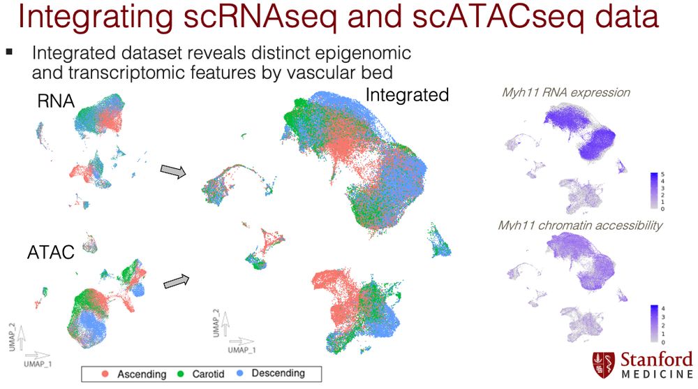
Gene regulatory network analysis through integrated RNA and ATAC datasets across cell types and vascular sites reveal cell type and vascular site specific GRNs, this highlighted ascending fibroblast specific MEOX1
10.09.2025 15:54 — 👍 1 🔁 0 💬 1 📌 0

Vascular site specific epigenomic patterns are distinct for SMCs, fibroblasts, as well as endothelial cells, but importantly not macrophage cells. While developmental TFs a enriched there are thousands of distinct enhancer elements across vascular sites
10.09.2025 15:54 — 👍 1 🔁 0 💬 1 📌 0

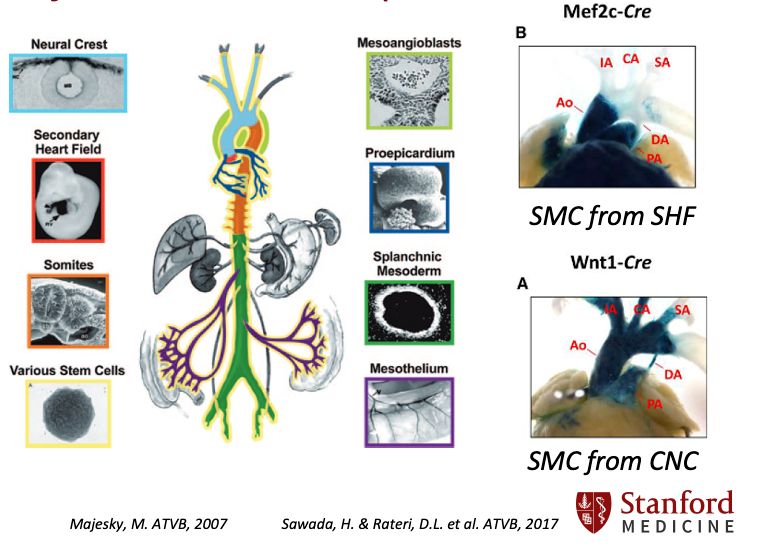
Many enhancers correspond to developmental origin and highlight specific developmental transcription factors such as HAND2, GATA4, and HOX family members, suggestive of an epigenetic 'memory' of developmental origin
10.09.2025 15:54 — 👍 1 🔁 0 💬 1 📌 0

In our work, by performing single cell RNA and ATAC sequencing across different vascular sites in mice, we reveal that the epigenomic landscape is distinct to not only cell type, but vascular site, defining vascular site specific enhancers
10.09.2025 15:54 — 👍 1 🔁 0 💬 1 📌 0

A critical element of complex genetics is that the majority of GWAS SNPs that influence disease regulate non-coding enhancer elements in the genome — where variants can influence disease risk through regulating cell type specific enhancers, but what about for the vasculature??
10.09.2025 15:54 — 👍 3 🔁 0 💬 1 📌 0

However we know that the genetic drivers of complex vascular traits vary based on vascular site, a beautiful example is the exceptional work by @jamespirruccello.com in 2022 showing the different genetic variants that impact ascending versus descending aortic dimension
10.09.2025 15:54 — 👍 2 🔁 0 💬 1 📌 0

Beautiful work in developmental biology going back decades has revealed that vascular diversity has a developmental basis, and that these vascular territories have distinct biology
10.09.2025 15:54 — 👍 1 🔁 0 💬 1 📌 0
Pronounce Bee-yo (🇫🇷)
Vascular biologist. Cat lover.
Asst Prof in Cardiovascular Medicine & Surgery at BWH/HMS in Boston
Formerly Pitt, UVA, Bordeaux.
Assist. Director of Bioinformatics | MGB Personalized Medicine
Investigator, Cardiovascular Research Center | Massachusetts General Hospital Member of the Faculty | Harvard Medical School
Interested in Cardiovascular Genetics and Personalized Medicine 🧬👨💻
RNAPII &Transcription Regulation -Chromatin Conformation & Nuclear Architecture - ESCs & Neuronal Differentiation - Nuclear Mechanotransduction - Cell Plasticity
UMG, Göttingen, Germany
We study the molecular and biophysical mechanisms underlying tissue morphogenesis @poldresden.bsky.social @tudresden.bsky.social @erc.europa.eu @embo.org
https://physics-of-life.tu-dresden.de/team/pol-groups/barriga
Chromatin and cancer and condensates. Lab Head in Discovery Oncology at Genentech.
AmyStrom.com
Postdoc @ Doudna Lab, UC Berkeley 🧬 | Chemical & Structural Biology | Immunity Mechanisms | CRISPR | RNA-Editing |
F32 NRSA | Berkeley Chancellor's Fellow
UC Davis Beal Lab Alum | Former CIRM, NIH F31 & NIH T32
Assistant Professor at U of Kentucky Medicine 🐾
Functional Genomics of Neural Activity and Animal Behavior at the single cell level
🧬genes->🚥pathways->🧠circuits->🐭behavior
European 🇪🇺 | #Berlin 🏠 | PhD student in medical systems biology 🧬 with @leifludwig.bsky.social | Cooking & mountains
Resident panhandler, http://trichelab.org/
Posts may contain trace quantities of blood🩸, chromatin 🧬, and stats 🧮
CV: https://scholar.google.com/citations?user=AOoIO74AAAAJ
Views expressed are my own (but for the right price they can be yours!)
Assistant Professor at the University of Michigan. Chromatin biology, transcription regulation, plant genomes.
https://marand-lab.github.io/
Genetic epidemiology postdoc at @stanford.edu
ICREA Research Professor. Evolution, Genetics, Neuroscience, Linguistic Cognition
Associate Professor at USC. Genetics/Stats/ML. Husband and father. GA➡️CA. He/him. Views are mine.
www.mancusolab.com
CS PhD Candidate at Stanford. Working at the intersection of Machine Learning, Regulatory Genomics, and Complex Disorders
Researcher - innate immunity, RNA sensors, viruses, inflammation, autoimmunity. Hudson Institute, Monash Uni, Melbourne.
Mum, T1D, tomato-grower, she/her
https://www.hudson.org.au/researcher-profile/natalia-sampaio/
Asst Prof of Molecular Cell Biology and Computational Biology at UC Berkeley
Building a new lab capturing close-ups of cells with electrons. lucaslab.science
Attempting to "dwell (somewhat) patiently in the variety and complexity of organisms."
Infectious diseases doctor, epidemiologist, gun injury prevention advocate, Stanford professor, and penguin enthusiast.


