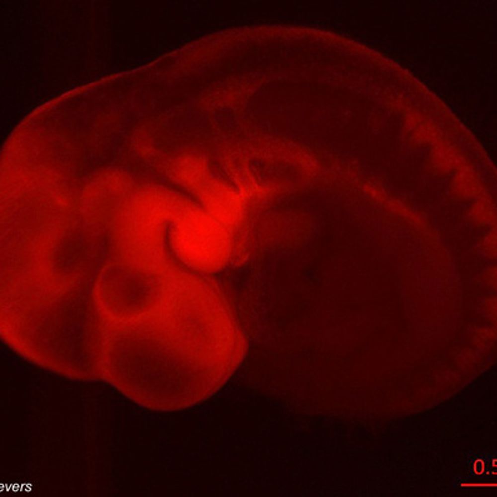
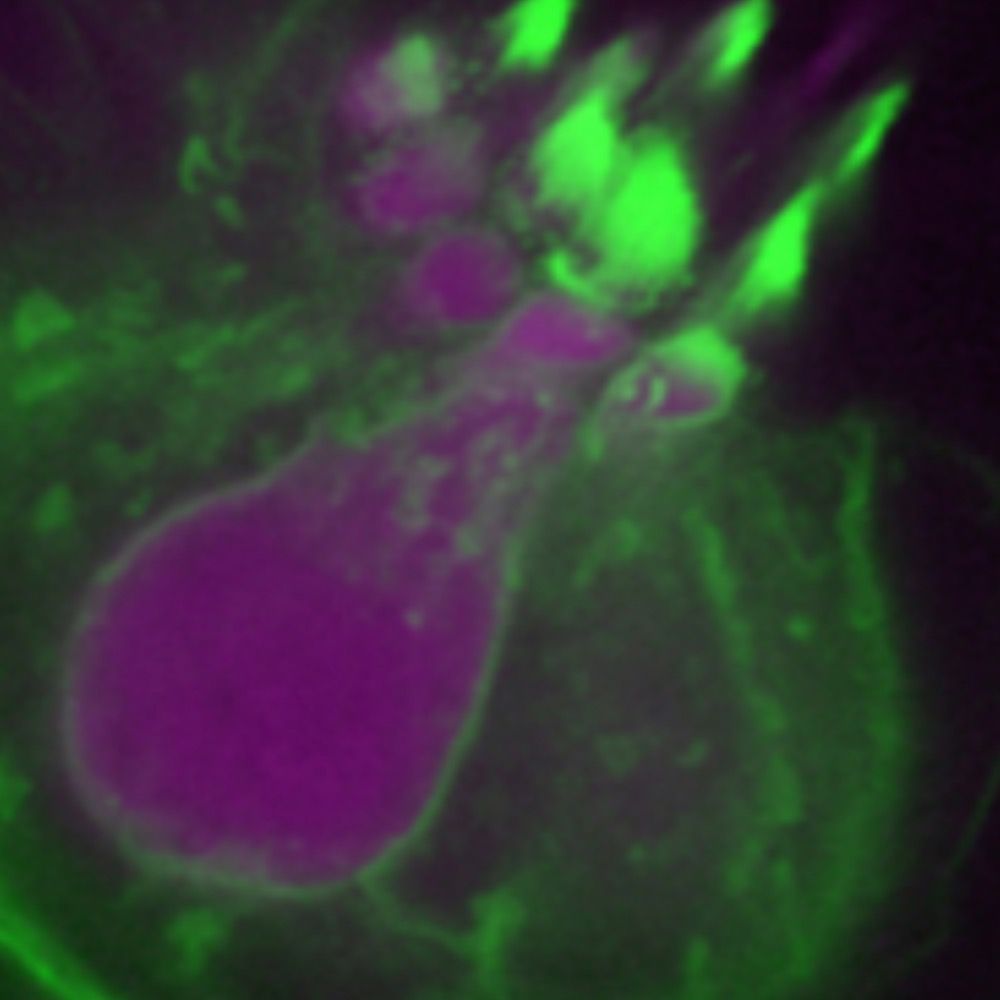
A widely used RNA assay labels the wrong molecules in several model organisms
After a routine experiment raised suspicions, Steinmetz group researchers joined forces with collaborators to highlight the limitations of a commonly used RNA labeling product.
New paper out in @bmc.springernature.com 🤩👏 A routine 5-ethynyl uridine (EU) RNA labeling experiment in a sea anemone turned into detective work for @malinkjosavik.bsky.social, @ktgarschall.bsky.social & @prhsteinmetz.bsky.social 🕵️♀️
29.01.2026 15:33 — 👍 20 🔁 9 💬 2 📌 2
Morphogenesis & Organogenesis!
Part 1 (full) in the comments 👇
Comment if you'd like to be added (regardless of age or career stage!)
Please post your own biology-related starter packs using #BioStarterPacks
🧬🔬🪰🐟🐁🌱
26.06.2025 21:40 — 👍 35 🔁 21 💬 18 📌 1
I would love to be added! This is great, thank you!
19.12.2025 18:37 — 👍 2 🔁 0 💬 1 📌 0

Confocal microscopy image of a juvenile sea star (Patiria miniata) viewed from the oral side. The animal has a five-armed, star-shaped body with a central nerve ring. The nervous system is labeled in green, forming radial nerve cords extending into each arm, and cell nuclei are labeled in red throughout the animal. The image appears against a black background and has a holiday-ornament-like appearance.
Felt a little festive at the microscope this morning for #FluorescenceFriday 🎄
Here’s the nervous system of a juvenile sea star ⭐️
Green = acetylated tubulin, red = nuclei
Happy holidays!
19.12.2025 17:32 — 👍 577 🔁 132 💬 10 📌 12

AI Is Inventing Academic Papers That Don't Exist -- And They're Being Cited in Real Journals
Academic articles from authors using large language model are creating an ecosystem of fake research that threatens human knowledge itself.
Academics and technologists are sounding the alarm about a growing crisis in scholarship as we know it: AI-generated citations of nonexistent papers that have infested real journals. Despite being fake, the sources are widely assumed to be authentic the more they appear in published literature.
17.12.2025 19:45 — 👍 945 🔁 498 💬 36 📌 160

watercolor of DNA gel
#ArtAdventCalendar Gel Electrophoresis in Green and Blue, watercolor, 2023 #sciart
12.12.2025 18:56 — 👍 138 🔁 33 💬 3 📌 0

From #amoeba to humans: new paper @natcomms.nature.com by the Grashoff group @sfb1348.bsky.social reveals evolutionary origin of animal cell adhesion and force transmission. Talin protein plays central role. See rdcu.be/eTFJR
@uni-muenster.de
08.12.2025 14:11 — 👍 8 🔁 6 💬 0 📌 1

Molecular basis for de novo thymus regeneration in a vertebrate, the axolotl
The molecular, cellular, and functional restoration of the axolotl thymus after de novo regeneration is described.
Can't believe my postdoc paper is finally out. Christmas came early this year, holy moly 🎄
Molecular basis for de novo thymus regeneration in a vertebrate, the axolotl | Science Immunology www.science.org/doi/10.1126/...
05.12.2025 21:17 — 👍 55 🔁 23 💬 4 📌 0

Join us for next week’s exciting VGZT session! 🎉
🗓️ Thursday, November 20th
⏰ 9:30 PST / 12:30 EST / 17:30 UTC / 17:30 GMT / 18:30 CET
Our speakers are
👉 Allison Kann (@apkann.bsky.social)
👉 Joana da Silva (on X: @joanamsilva14)
See you there 👋
14.11.2025 08:43 — 👍 7 🔁 8 💬 0 📌 1

This image by MDI Bio Lab's Travis Carney is a #drosophila larval brain. Neural stem cells and neurons are marked, including axons that project into the brain. The flare in the center of each lobe is part of a learning and memory center in flies.
ZEISS Microscopy #microscopymonday 🧪 🤝
10.11.2025 15:01 — 👍 7 🔁 5 💬 0 📌 0
PAPER OUT ✨ What if you could use your microscope as a 3D printer? Prototype microfluidics in-house, <5$ in material costs per chip. From idea to experiment within a day. Now published in Lab-on-a-Chip (open-access): doi.org/10.1039/D5LC...
16.07.2025 11:51 — 👍 91 🔁 22 💬 4 📌 4

As spooky season is upon us 🎃, we have cells that look like spider webs for #FluorescenceFriday 🧪🔬
24.10.2025 19:03 — 👍 70 🔁 10 💬 1 📌 0
Happy to share that this work is now published in @biophysj.bsky.social! doi.org/10.1016/j.bp...
23.10.2025 16:30 — 👍 25 🔁 6 💬 1 📌 0
A toolkit for testing membrane-localising tags across species
Transgenic markers and tools have revolutionised how we study cells and developing organisms. Some of the elements needed to construct those tools are universally applicable (e.g. fluorescent proteins...
How can we see the cells that make up a living organism? Membrane-localising tags can drive fluorescent proteins to the cell's outer membrane, making their outlines visible. But the tags don't work well in all organisms. How do you find one for your species of interest? 🧵
Check our latest preprint
23.10.2025 08:16 — 👍 87 🔁 41 💬 5 📌 5
One of the coolest projects I've seen in years - huge congrats to Çağrı and the McKinley team!
10.10.2025 15:09 — 👍 7 🔁 1 💬 1 📌 0
🚨 My lab is hiring at all levels!
Interested in animal origins & evolutionary cell biology?
I'm recruiting a postdoc, PhD students & a research assistant to study the molecular evolution of cell adhesion using marine invertebrates + comparative genomics.
🔗: clarkelab.com/join/
Please repost!
08.10.2025 17:49 — 👍 90 🔁 68 💬 1 📌 1

RhoA (blue) and the actin cytoskeleton (magenta) are shown in a set of primary microglia.
For #FluorescenceFriday, RhoA (blue) and the actin cytoskeleton (magenta) are shown in a set of primary microglia 🔬 #Neuroscience #Microscopy
03.10.2025 13:07 — 👍 83 🔁 16 💬 4 📌 1
Absolutely gorgeous work from Kate - I loved reading the full paper!
02.10.2025 15:43 — 👍 2 🔁 0 💬 0 📌 0
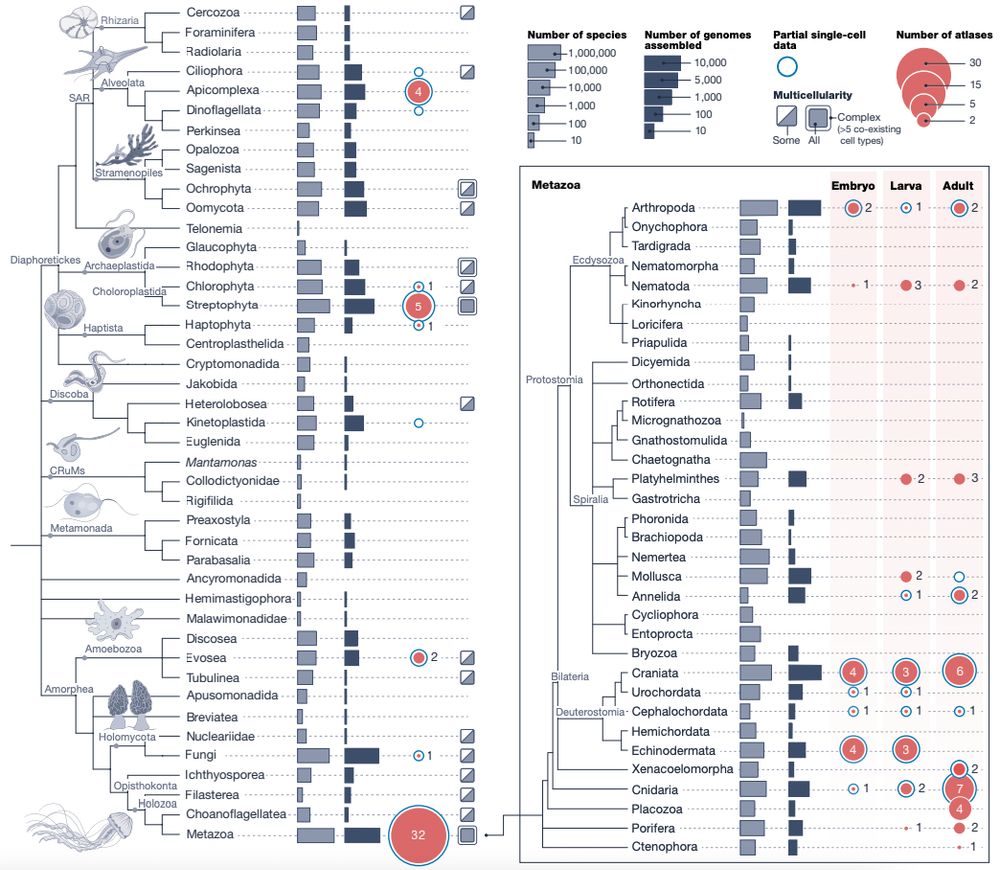
Happy to share the Biodiversity Cell Atlas white paper, out today in @nature.com. We look at the possibilities, challenges, and potential impacts of molecularly mapping cells across the tree of life.
www.nature.com/articles/s41...
24.09.2025 15:12 — 👍 228 🔁 106 💬 4 📌 10

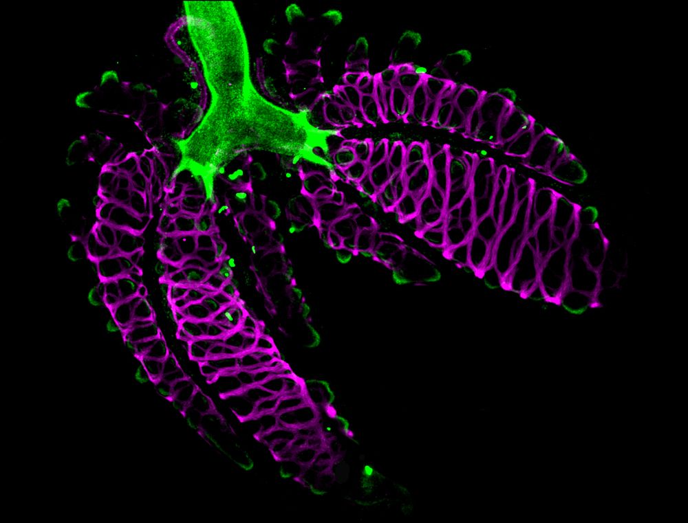
brightfield images of three lizard embryos of approximately the same developmental stage. Below each embryo image is an immunofluorescence image labeling E-cadherin (green) and alpha-smooth muscle actin (magenta) of their developing lungs
New preprint from some of my postdoc work on lungs! Co-led with Kaleb Hill, we studied smooth muscle and epithelial development in lizard lungs. Stay tuned for more!
www.biorxiv.org/content/10.1...
05.09.2025 18:12 — 👍 22 🔁 10 💬 2 📌 1

Peripheral muscle fibers (pseudocolored in magenta) and nuclei (in gray)
Today's #FluorescenceFriday is featuring the peripheral muscle of Hofstenia miamia 💪
29.08.2025 14:27 — 👍 65 🔁 9 💬 0 📌 0

Cells can form patterns within themselves just like embryos do. How? Connie Yan's new preprint shows how the anterior-posterior cytoskeleton pattern in Stentor is dictated by regionalized scaffolding proteins
www.biorxiv.org/content/10.1...
19.08.2025 08:41 — 👍 142 🔁 35 💬 3 📌 5

Screenshot of Essay from Martin Schwartz on 'Why would anyone want to be a scientist'. An anniversary article from The Company of Biologists published in Journal of Cell Science.
The first few lines are: It is difficult to fathom why anyone intelligent enough to be a scientist would actually choose to be one. Doing good science requires the utmost exertion of body, mind and spirit, yet is consistently filled with failure and rejection. But, strange even to myself, I not only don't question the unfavorable risk-to-reward ratio but consider myself astonishingly lucky to be a scientist. There are three fundamental pleasures that have sustained me through 50 years of this madness.
Why would anyone want to be a scientist?
Check out our new Essay from Martin Schwartz: journals.biologists.com/jcs/article/...
15.08.2025 13:19 — 👍 81 🔁 46 💬 2 📌 8
Latest paper elifesciences.org/articles/107... closes an important cycle in our efforts to study regeneration: week-long recordings allow us to observe the behaviour of cells during the entire course of regeneration in a crustacean leg – bright objects in movie are fluorescent nuclei of cells. 1/6
08.08.2025 17:39 — 👍 142 🔁 50 💬 2 📌 3
✨Development meets design in this embryonic chameleon lung 🫁 🦎. Smooth muscle swirls 🟣 and branching tips and cartilage 🟢 come together in reptilian lung morphogenesis. Image taken by @drkatiegoodwin.bsky.social 🔬🧪
#FluorescenceFriday #DevBio
08.08.2025 13:11 — 👍 51 🔁 14 💬 0 📌 1
Fluorescent embryo showing the developing epidermis, pseudo-colored with the ICA LUT in FIJI
Our transgenic acoel embryos are beautifully mosaic 😍 showing off the developing epidermis for #FluorescenceFriday
08.08.2025 13:57 — 👍 28 🔁 5 💬 0 📌 0
Congratulations Tim!! So exciting!
03.07.2025 15:59 — 👍 0 🔁 0 💬 1 📌 0
🇲🇽 PhD Student @MPIforBI / UniGoe interested in ethology, neural computations and evolution. NSB-MBL '25
Post doc in the Burkhardt group at the Michael Sars Centre @UiB | Embryology 2024 @MBLScience | Interested in nervous system evolution (mostly invertebrates)
PhD candidate in molecular evolution @UChicago
manyuanlonglab.uchicago.edu
Neuroscientist at VIB KULeuven
Exploring Hox genes regulation in Stem Cell-Based Embryo Models at @college-de-france.fr | Duboule Lab | CDSN PhD Fellowship
postdoctoral researcher at the MPI for biological intelligence I development & evolution of the brain I interactions of transcription factors
Group of Adrien Hallou @kiroxford.bsky.social @ox.ac.uk
Biophysics & spatial biology of cell fate decisions & tissue dynamics
Alumnus @cam.ac.uk & @normalesup.bsky.social
Franco-British Young Leader 2024 🇨🇵🇬🇧
Neural development enthusiast, from neurulation to cell differentiation. Chick and zebrafish embryos 🐣🐟🔬. Facultad de Ciencias, Universidad de la República, Uruguay. @fzolessi.bsky.social lab #DevelopmentalBiology #Neuroepithelium #Retina #CellPolarity 🚲🇺🇾
Interested in mechanobiology, morphogenesis, organisers and synthetic embryo models.
Research Fellow in Mongera lab UCL | PhD in Mayor and Charras labs | YEN Committee
Co-director of MoPED (Mechanisms of Paracrine & Endocrine Disorders) lab in Marseille 🇫🇷 on cell signaling & determination of self & beyond during neural crest differentiation. Personal account.
all things cytoskeleton - nuclear actin dynamics and genome - nucleoskeleton and nuclear organisation - cancer cell invasion and pharmacology
@University of Freiburg
Assc Prof @pennmedicine.bsky.social.
Co-Director, McKay Orthopaedic Research Labs.
Mechanobiology of development & regeneration.
Prov. 25:2. 🦛
Philadelphia, PA
Living a life of troubleshooting🫠
🐶Univ. of Washington, JSPS Overseas Postdoc fellow ←🌱Univ. of Tsukuba, Japan.
Arata Wakimoto / 脇本 新
JP/EN
#StemCells, #DevBio, #EvoDevo
Love☕ #Coffee and 👾 #Games
Dr Tim Grocott's research group, University of East Anglia UK. 🐣 embryology & new adventures in human development via stem cells. Modelling in silico/vitro. Fascinated by signalling networks, patterning, all things early development and the eye.
PhD candidate @MSarsCentre / Computational biology / Development biology / Image analysis / Data diver
PhD - Senior Research Assistant in Stapornwongkul Lab (IMBA, Vienna).
Cell and developmental biologist.
Passionate bioimager and photographer.
Hi, I am a scientist working on epithelial Morphogenesis @IBDM Marseille. Passionate about Development, Shape, Evolution, and turning pretty pictures into numbers
Developmental cell biologist exploring epithelial morphogenesis, tube formation, tricellular junctions, and more. Views are my own.
www.luschnig.uni-muenster.de/
Postdoc | Institue for Psychedelics and Neurotherapeutic | Olson Lab | UC Davis
PhD | CIRM fellow | Cerebral Organoids | Haussler-Salama Lab | UC Santa Cruz
hair cell development, death and regeneration
https://faculty.washington.edu/draible/