A cell finding its way through the matrix, imaged with @joycemeiri.bsky.social on LLS.
19.09.2025 10:38 — 👍 34 🔁 13 💬 1 📌 1
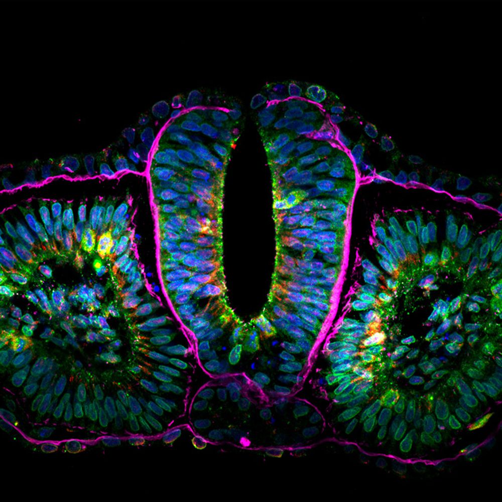
Our new preprint is about the airway epithelium microtubule network, cilia, basal body protein composition, averaging of volumetric fluorescence data, and expansion microscopy.
Four years of very hard work from our very talented Emma van Grinsven. www.biorxiv.org/content/10.1... (1/N)
05.09.2025 15:37 — 👍 41 🔁 16 💬 2 📌 1

Our September issue is here! rupress.org/jcb/issue/22...
Airyscan super-resolution immunofluorescence image of HeLa cells stained for two types of intermediate filaments: vimentin (magenta) & keratin-8 (cyan). From Pasolli, @joycemeiri.bsky.social, Akhmanova et al. rupress.org/jcb/article/...
01.09.2025 14:03 — 👍 14 🔁 5 💬 0 📌 0
All the opto-vimentin plasmids are now available on Addgene: www.addgene.org/Anna_Akhmano...
08.08.2025 15:51 — 👍 11 🔁 3 💬 0 📌 0
Radial microtubules in cyan and non-radial microtubules in yellow.
18.07.2025 10:06 — 👍 3 🔁 0 💬 0 📌 0
Optogenetic and chemical genetic tools for rapid repositioning of #vimentin intermediate filaments. New tools developed by Milena Pasolli, Joyce Meiring @joycemeiri.bsky.social, Anna Akhmanova and colleagues @utrechtuniversity.bsky.social: rupress.org/jcb/article/...
@gijsjekoenderink.bsky.social
15.07.2025 13:45 — 👍 18 🔁 3 💬 0 📌 0
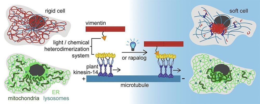
New: Pasolli, Meiring, Akhmanova et al. @utrechtuniversity.bsky.social describe tools to acutely relocate #vimentin intermediate filaments by inducibly coupling them to microtubule motors. rupress.org/jcb/article/...
@joycemeiri.bsky.social @gijsjekoenderink.bsky.social
#Technology #Cytoskeleton
08.07.2025 16:01 — 👍 13 🔁 5 💬 0 📌 0
Here is an example of the reversibility. The first 30 min the cell is pulsed with blue light (when blue dot is shown) causing the tagged vimentin to associate with - end oriented kinesins that pull the vimentin to the cell centre. Then blue light pulsing is stopped and vimentin redistributes.
08.07.2025 15:42 — 👍 4 🔁 1 💬 0 📌 0

Excited to share that our fast & reversible chemical and optogenetic vimentin pulling tools are now published in JCB! 🥳
We show a subset of organelles are displaced with vimentin pulling to the cell centre and cells become much softer despite little or no effect on actin/MTs
doi.org/10.1083/jcb....
08.07.2025 15:34 — 👍 24 🔁 7 💬 1 📌 1
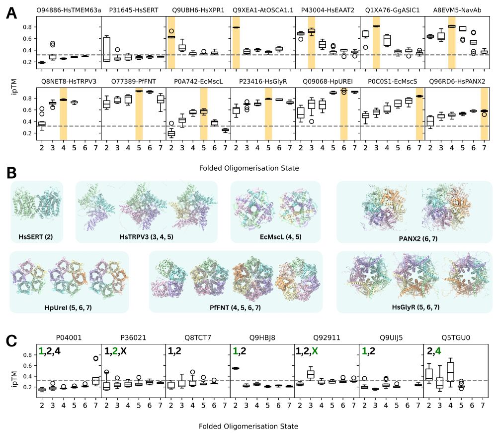
Thrilled to share our latest preprint from @yiechanglin.bsky.social & @ciarawallis.bsky.social showing that AF2-Multimer and AF3 can quickly & accurately predict the oligomeric states & structures of proteins (& maybe can identify multiple biologically relevant oligomeric states 👀)
tiny.cc/oligomers
16.03.2025 06:37 — 👍 36 🔁 8 💬 1 📌 1
Don't forget to register, abstract submission deadline is next week March 14. 😉
07.03.2025 11:08 — 👍 1 🔁 0 💬 0 📌 0
Congratulations!! 🥳🥂
27.02.2025 19:51 — 👍 1 🔁 0 💬 0 📌 0
Is this an animated Jackson Pollock painting or live cell tracking movie? 😍
19.02.2025 16:12 — 👍 5 🔁 0 💬 0 📌 0

Come join us in Utrecht for some cutting edge #microscopy and 3D cell biology!! #3Dcellbio
29.01.2025 08:24 — 👍 30 🔁 10 💬 1 📌 2
Hi! I was wondering if anyone had successfully cultured happy HEK293T cells in defined serum alternatives? If so, what did you use?
27.11.2024 05:36 — 👍 3 🔁 5 💬 0 📌 0
Excited to share our new pre-print showcasing tools (chemical & optogenetic) for repositioning Vimentin in cells! Movie shows ER (Halo-KDEL) sheets being relocalized together with vimentin (Vim) upon local vimentin pulling in U2OS. @gijsjekoenderink.bsky.social
www.biorxiv.org/content/10.1...
22.11.2024 13:58 — 👍 14 🔁 0 💬 0 📌 0
Time to shine for intermediate filaments! An optogenetic vimentin perturbation strategy reveals its role for ER and mitochondria positioning (but no effect on actin or microtubules patterns) @joycemeiri.bsky.social
www.biorxiv.org/content/10.1...
21.11.2024 22:40 — 👍 33 🔁 9 💬 1 📌 0
CAMSAP3 stretches (microtubule -end binding protein) are shown in yellow. They move towards the Soma in developing neurons.
20.11.2024 07:56 — 👍 4 🔁 1 💬 1 📌 0
We like the Cytochrome C from BD Pharmingen cat#556432
20.11.2024 05:53 — 👍 2 🔁 0 💬 0 📌 0
This video shows the smallest area I've managed to sever inside a U2OS cell. The goal for this experiment was to sever microtubules around a subset of the Golgi (labelled here in cyan, together with exocytic vesicles) to see what would happen to it's morphology/localization.
19.11.2024 15:37 — 👍 0 🔁 0 💬 0 📌 0
However in a neuron it could be a smaller area still, because diffusion is slower in neurites due to their morphology. I haven't tried to see how small the area could be in neurons though.
19.11.2024 13:41 — 👍 1 🔁 0 💬 1 📌 0
Good question, I would estimate an area of around 10 um^2.
19.11.2024 13:22 — 👍 1 🔁 0 💬 0 📌 0
Hi! My name is Joyce and I love building and using tools to manipulate the #cytoskeleton of cells to better understand their function. 🔬
Below you can see my movie of #opto-katanin ✂️(SspB-mCh-p60) being used to locally disassemble #microtubules (labelled by SiR-tubulin) inside a neuron.
19.11.2024 07:44 — 👍 104 🔁 18 💬 7 📌 0
Comparative developmental biology, regeneration, non-conventional model organisms, live imaging; see www.averof-lab.org
@cshlnews.bsky.social postdoc with @hannahvmeyer.bsky.social and Saket Navlakha • How your T cells know it's you • Develops immuno tools named after Gotham characters: github.com/meyer-lab-cshl/BATMAN 🦇 • they/he 🏳️🌈🏳️⚧️
Microscopes, desmosomes, cell adhesion, endocytosis, cell mechanics at UAB
Cytoskeketal cell biologist
Associate Professor in Cell and Molecular Physiology at Loyola University Stritch School of Medicine.
Opinions are my own or sampled from other people who are smarter or funnier than I.
Postdoc with Gaia Pigino @ Human Technopole. Previously PhD student with Andrew Carter.
Journal of Cell Biology publishes peer-reviewed research on all aspects of cellular structure and function. Published by Rockefeller University Press @rupress.org
🌐 https://rupress.org/jcb
Biochemist excited about fluorescent proteins, multiplexed imaging and SOFI.
PhD at Université Paris Saclay, currently postdoc in @dedeckerlab.bsky.social at KU Leuven.
Big fan of science communication.
PhD at the University of Amsterdam. Microscopy enthusiast 🔬, cytoskeleton admirer 🧫, hopeful sensor builder. Mostly just a biologist finding his way.
Neuroscientist at the Paris Brain Institute / www.dejuansanzlab.org / ERC / FENS-Kavli Scholar / Young Academy of Spain / CNRS
WIMR is a leading Australian medical research institute delivering groundbreaking discoveries to better prevent, diagnose, treat and cure some of the most serious global health issues.
POTATOMap|Lysosomes|Neuronal Organelle|UU PhD Candidate|Farías lab|Utrecht University|Cell Biology|MPhil. Life Science HKUST
CNS regeneration lab, King's College London, Wolfson SPaRC. We use neuronal cell biology to study axon regeneration, identifying and testing new therapies for eye disease and spinal cord injury.
Inserm researcher: Neuroscience, Imaging, cell biology, super-resolution. Sci. head of NeurImag facility in Paris
I am a Software Architect for AI Solutions, Python Fan, Microscopist Image Analyst and really like Skiing.
Science is like magic but real.
Research Development | Grant Writing | ex-Scientist (Cell Division Lab, Mitosis, Cancer, Microscopy) | Bodyboarding | Gold Coast 🇦🇺 |
AndrewBurgessPhD.com
Opinions are my own
Super Talented Wife: vrmorrison.com rosemorrisonartist.com
#EmmyNoether Lab of Hans Maric @uni-wuerzburg.de
#ChemBio #Peptides #PNA #Microarrays #ChemicalProbes pharmacological targeting of #PPI #IDR #RNA
http://MaricLab.com
https://www.uni-wuerzburg.de/en/rvz/research-groups/maric-group/
http://bit.ly/14S4Z8k.