🚨 New publication alert:
📢 “Sketchpose: Learning to Segment Cells with Partial Annotations.”
🖊️ @ccazorla31.bsky.social, N Munier, R Morin, P Weiss.
⬇️
28.08.2025 13:42 — 👍 1 🔁 1 💬 1 📌 0

#Sketchpose is out in @melbajournal.bsky.social !
Go read it if you are interested in training cell segmentation models using few partial annotations 😀
25.08.2025 09:28 — 👍 4 🔁 2 💬 0 📌 0
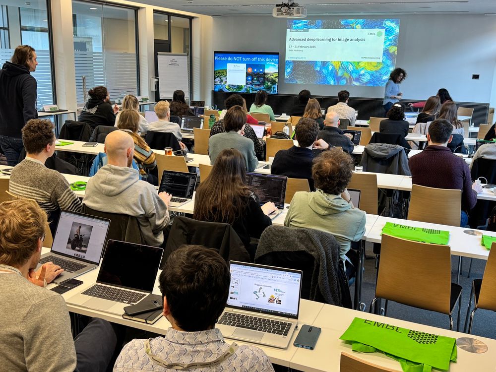
‼️🔬 We start!!! 🎉🤖
Super exciting!!! After years of teaching courses for #DeepLearning for Microscopy and Life Science application, we start today our first “advanced course” @embl.org!
#EMBLDeepLearning
17.02.2025 08:37 — 👍 107 🔁 15 💬 2 📌 2
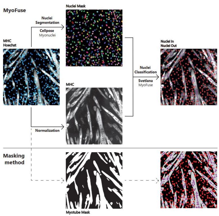
Thrilled to share MyoFuse, an AI-based workflow for automated skeletal muscle cell fusion quantification! 🥳
This is a collaboration with Benjamin Lair and Cedric Moro (I2MC, Toulouse), improving the Fusion Index (FI) measurement.
Preprint: www.biorxiv.org/content/10.1...
#AI #Svetlana #Myofuse
18.02.2025 10:03 — 👍 11 🔁 5 💬 0 📌 0
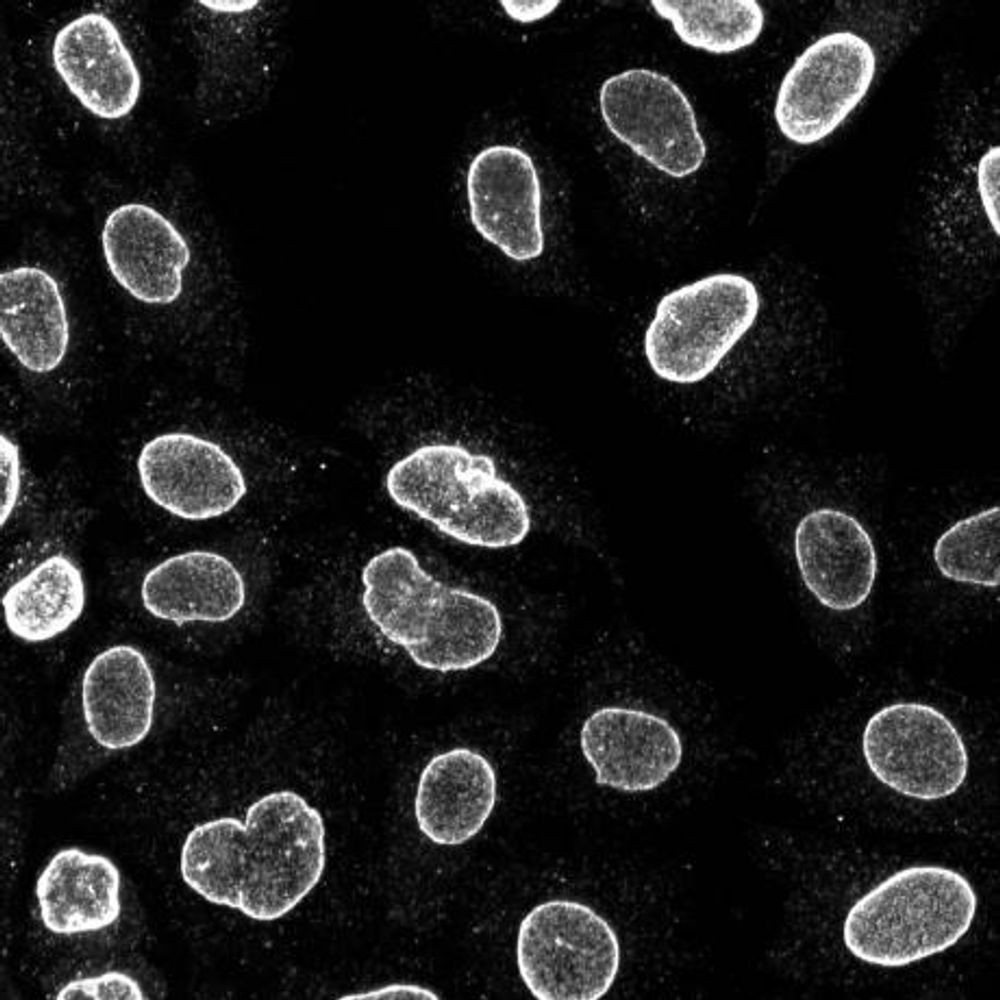
🚨 New segmentation model alert! 🚨🥳
You can finally use InstanSeg in our #BioImageAnalysis software #CellACDC!
InstanSeg is a fast and multi-channel cell segmentation model by Pete Bankhead, Thibaut Goldsborough, & co. The multi-channel ability is very interesting!
22.12.2024 19:07 — 👍 5 🔁 2 💬 1 📌 0
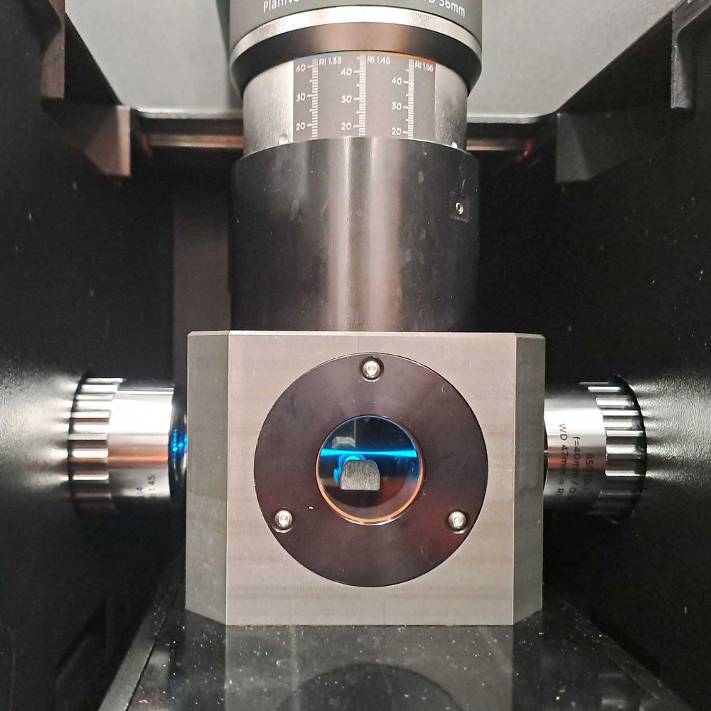
Interested in SMCs, microscopy, biophysics, hiking, ...
working at MPI for biophysics, FFM
Imaging Scientist at Facility for Imaging by Light Microscopy (FILM) @imperialcollegeldn.bsky.social
Previously FILM facility manager.
Dad, Cell biologist, PhD/postdoc
@uniofcam.bsky.social.
Opinions my own.
PhD student at ENS PSL; studying the role of cytoskeleton in shaping the nuclear architecture during ependymal cell differentiation. #mechanotransduction #devbio #microfluidics
Microscopist and image analyst at GIGA - Uliege, Belgium 🇧🇪 passionate by life sciences, geeky father of 3
Lead Editor: #CRBIOTECH & #ExplorDHT | PI: PaDiH & LBI-DHPS | Prof of IGAB-PAS | Leader: #DHPSP & #INPST | Expert Consultant in Biotech & Science Communication
🔗Web: https://digitalpatientsafety.com/atanas-g-atanasov/
Cellular Biologist, Microscopy, Image analysis, Geek, Dad
Research group lead by @henriqueslab.bsky.social at @itqbnova.bsky.social
Combining #AI with #superresolutionimaging to push the boundaries of optical cell biology #SMLM #SRRF
Chercheur, vulgarisateur et youtubeur sur https://www.youtube.com/@Fouloscopie
Des repères clairs et de la culture pour tous !
Associate Prof at Abo Akademi University, Turku, Finland.
Cell biologist interested in microscopy and image analysis. My opinions are my own.
cellmig.org
CNRS researcher
// 🔬 microscopy // 🗜️ microfabrication // 🧫 bioengineering // ☯️ cell self-organisation //
http://biof-lab.org/
Microscopist & Python developer at Harvard Med. Creator @fpbase.org
I am an n-dimensional image viewer for Python
🌉 bridged from ⁂ https://fosstodon.org/@napari, follow @ap.brid.gy to interact
The Imaging Core Facility of the Biozentrum is a technology platform for the use of light microscopes and users support.
BioImage Analyst
Head of BioImage Informatics, SciLifeLab
NBIS, Sweden
https://www.scilifelab.se/units/bioimage-informatics/
Group leader @ Uni Göttingen for Computational Cell Analytics. Working on AI for biology and medicine with a focus on microscopy image analysis with deep learning.
https://user.informatik.uni-goettingen.de/~pape41/
Artificial Intelligence For Image Data Analysis in The Life Sciences, funded by Horizon Europe, created to serve YOU!
Bio-image Analyst, Computer scientist, and plenty of other things that end in «-ist» @pasteur.fr