Powered by MHR
We are recruiting a postdoc to join a Wellcome Trust-funded project at LSI-Exeter, investigating the role of neural-pancreas dynamics in maintaining glucose homeostasis. 🐟🔬🧠
@lsiexeter.bsky.social #zebrafish #islet #postdocjobs
jobs.exeter.ac.uk/hrpr_webrecr...
Please repost 🙏
04.02.2026 17:33 — 👍 7 🔁 3 💬 1 📌 1

Promotional graphic announcing that scientific submissions for EASD 2026 are open. Text highlights the dates “28 September-02 October” and the location “Milan, Italy.” A message invites researchers to submit abstracts to share their work with the global diabetes community. The design features red and white brush-stroke panels and a stylised skyline of Milan on the right, with “EASD 2026 Milan” and “easd.org” at the bottom.
🎯 Started preparing your #abstract submission for the 62nd EASD Annual Meeting? This is your sign to get started & share your #diabetes #research with the global community in Milan 🇮🇹 from 28 Sept - 02 Oct 2026.
🗓️ Deadline: 1 Apr 2026 | 🕛 18:00 CEST
Submit via MyEASD 👉 www.easd.org/annual-meeti...
03.02.2026 10:09 — 👍 10 🔁 7 💬 0 📌 0

Call for Applications graphic for the EFSD Rising Star Fellowship Programme and EASD Rising Star Symposium. Text encourages young researchers in Europe to develop innovative diabetes research. Supported by Novo Nordisk through a donation. Includes EFSD logo, website europeandiabetesfoundation.org, abstract blue and green shapes, and an image of a researcher using a microscope.
📢🚨 Don’t miss this: Apply for the #EASD Rising Star Symposium and #EFSD Rising Star Fellowship Programme!
Eligibility: EASD members, within 7 years of PhD/MSc/MD
⏰ Deadline: 17 Feb 2026 | 12:00 CET
👉 Apply: www.europeandiabetesfoundation.org/programmes/p...
Supported by Novo Nordisk
#diabetes
05.02.2026 10:01 — 👍 1 🔁 1 💬 0 📌 0
Would you like to participate in the peer review process at Communications Biology? Please fill out this form to express your interest to act as a reviewer📝 We particularly encourage early-career researchers and applicants from underrepresented groups in research. forms.office.com/e/fv22f9aRt5
06.08.2025 16:41 — 👍 8 🔁 9 💬 0 📌 0

Fig. 1. Animal cap assay and sandwich method as in vitro induction systems.
In amphibians, a blastocoel cavity clearly forms inside the animal hemisphere during the blastula and early gastrula stages. The cap-like portion lining the roof of the blastocoel cavity is the animal cap. This region consists of a sheet of pluripotent cells, organized into one or several layers. In the animal cap assay, the animal cap was treated with a physiological saline solution containing inducing factors and then cultured. Depending on the type, concentration, and duration of exposure to the inducing factors, animal caps can differentiate into various cell types. In contrast, the sandwich method, involves culturing the inducer source in between two animal caps. In this technique, the sources of induction can include the dorsal lip of the blastopore (organizer), adult tissues, pelletized soluble factors, or animal caps pretreated with soluble factors. In this figure, activin is used as an example of an inducing factor.
![Fig. 12. Summary of the in vitro induction system using activin as an inducing factor.
This in vitro induction system utilizes activin and retinoic acid as inducing factors to treat animal caps, employing techniques such as animal cap assay, dissociation/reaggregation protocol, and the sandwich method. By applying these methods, various levels of self-organization can be replicated and controlled in vitro, ranging from lower-order cell differentiation to higher-order tissue differentiation, organogenesis, and even the formation of fundamental body plans. Abbreviations: Dorsal [D], ventral [V], and retinoic acid [RA].](https://cdn.bsky.app/img/feed_thumbnail/plain/did:plc:cnykzbo2impysrf6a6scy6oh/bafkreiarcsezsnfoj4xmlow6thcxsfy6wsfxfujek53lgt6wsw2k2wshqe@jpeg)
Fig. 12. Summary of the in vitro induction system using activin as an inducing factor.
This in vitro induction system utilizes activin and retinoic acid as inducing factors to treat animal caps, employing techniques such as animal cap assay, dissociation/reaggregation protocol, and the sandwich method. By applying these methods, various levels of self-organization can be replicated and controlled in vitro, ranging from lower-order cell differentiation to higher-order tissue differentiation, organogenesis, and even the formation of fundamental body plans. Abbreviations: Dorsal [D], ventral [V], and retinoic acid [RA].
![Fig. 11. Formation of embryoids by artificial activin concentration gradients.
To create embryoids, animal caps were prepared through treatment with low (0.5–1 ng/ml), intermediate (5–10 ng/ml), or high (50–100 ng/ml) concentrations of activin. These three types of activin-treated animal caps were then sequentially arranged and cultured with untreated animal caps. After 3 days of culture, embryoids with distinct head and trunk-tail structures were formed (A). Histological sections revealed differentiation into head tissues, such as the cement gland [cg] and eyes, and trunk-tail tissues including the ear vesicle [ev], brain [br], notochord [not], muscle [mus], and gut (B). When newt embryos are used in similar combination cultures, neural plate structures forming the brain [white arrow] and axial structures forming the trunk-tail regions [black arrow] are sometimes observed (C).](https://cdn.bsky.app/img/feed_thumbnail/plain/did:plc:cnykzbo2impysrf6a6scy6oh/bafkreig36tdo3ewakfxhn66uzs2kfqlkjjoebdcfh5rzy3ubxkfnn7t5iu@jpeg)
Fig. 11. Formation of embryoids by artificial activin concentration gradients.
To create embryoids, animal caps were prepared through treatment with low (0.5–1 ng/ml), intermediate (5–10 ng/ml), or high (50–100 ng/ml) concentrations of activin. These three types of activin-treated animal caps were then sequentially arranged and cultured with untreated animal caps. After 3 days of culture, embryoids with distinct head and trunk-tail structures were formed (A). Histological sections revealed differentiation into head tissues, such as the cement gland [cg] and eyes, and trunk-tail tissues including the ear vesicle [ev], brain [br], notochord [not], muscle [mus], and gut (B). When newt embryos are used in similar combination cultures, neural plate structures forming the brain [white arrow] and axial structures forming the trunk-tail regions [black arrow] are sometimes observed (C).
![Fig. 7. In vitro heart formation and in vivo transplantation experiment.
When treated with a high concentration of activin, the animal caps of Xenopus embryos did not differentiate into heart tissue. However, if the animal cap dissociates into individual cells before activin treatment and then reaggregates, it forms a beating heart [arrow] with 100 % efficiency (A). This heart expresses differentiation marker genes, such as Nkx2.5, GATA-4, Tbx5, MHCα, TnIc (cardiac troponin I), and ANF, none of which are expressed in an animal cap treated with activin alone, without dissociation/reaggregation (B). Electron microscopy reveals the presence of intercalated discs [id] specific to the cardiac muscle, along with visible mitochondria [m] and Z-bands [z] (C). When the reaggregated heart tissue is orthotopically transplanted into the cardiac primordium of a neurula-stage embryo, it integrates without rejection and continues to beat (D), although it does not persist through host metamorphosis. In contrast, when the reaggregated tissue is ectopically transplanted into the ventral region of the neurula, it begins to beat synchronously with the host heart and gradually reddens as it initiates blood circulation (E).](https://cdn.bsky.app/img/feed_thumbnail/plain/did:plc:cnykzbo2impysrf6a6scy6oh/bafkreig77lwzs67jdcjwi2gjsany3shyoa5vj2dpetburc3pegwxeb45a4@jpeg)
Fig. 7. In vitro heart formation and in vivo transplantation experiment.
When treated with a high concentration of activin, the animal caps of Xenopus embryos did not differentiate into heart tissue. However, if the animal cap dissociates into individual cells before activin treatment and then reaggregates, it forms a beating heart [arrow] with 100 % efficiency (A). This heart expresses differentiation marker genes, such as Nkx2.5, GATA-4, Tbx5, MHCα, TnIc (cardiac troponin I), and ANF, none of which are expressed in an animal cap treated with activin alone, without dissociation/reaggregation (B). Electron microscopy reveals the presence of intercalated discs [id] specific to the cardiac muscle, along with visible mitochondria [m] and Z-bands [z] (C). When the reaggregated heart tissue is orthotopically transplanted into the cardiac primordium of a neurula-stage embryo, it integrates without rejection and continues to beat (D), although it does not persist through host metamorphosis. In contrast, when the reaggregated tissue is ectopically transplanted into the ventral region of the neurula, it begins to beat synchronously with the host heart and gradually reddens as it initiates blood circulation (E).
A fascinating review on the role of Activin in organ induction. Isn't it wild that in Xenopus embryos, a piece of the animal cap can be induced with Activin at different concentrations and buffers to form the ❤️, kidney, the pancreas, head, tail, and even a whole embryoid 🤯:
doi.org/10.1016/j.cd...
31.01.2026 18:39 — 👍 23 🔁 8 💬 0 📌 2

Explore emerging research with field leaders Keystone Symposia #IsletBiology & #Diabetes : #BetaCell Compensation, Failure & Recovery, this March in Breckenridge! #KSDiabetes26
For more info:
➡️ https://hubs.la/Q03X7Ckz0
21.01.2026 16:01 — 👍 6 🔁 3 💬 0 📌 0

That 5th sample...
#lama #portal #aperturesciencesentryturret #thinkingwithportals
23.01.2026 22:00 — 👍 2 🔁 0 💬 0 📌 0

Nicolae Paulescu - Wikipedia
well, Nicolae Paulescu isolated insulin, although he called it pancreine and even try to patent it in 1916 - but Banting, Best and Macleod got it to work in humans. Probably it matters who gets the best outcome of research more than who had the idea first…
en.wikipedia.org/wiki/Nicolae...
18.01.2026 16:31 — 👍 2 🔁 0 💬 1 📌 0
Not sure, it takes a lot to keep up with being scientist, but I would say that some people truly dislike other scientists…
18.01.2026 16:17 — 👍 0 🔁 0 💬 0 📌 0

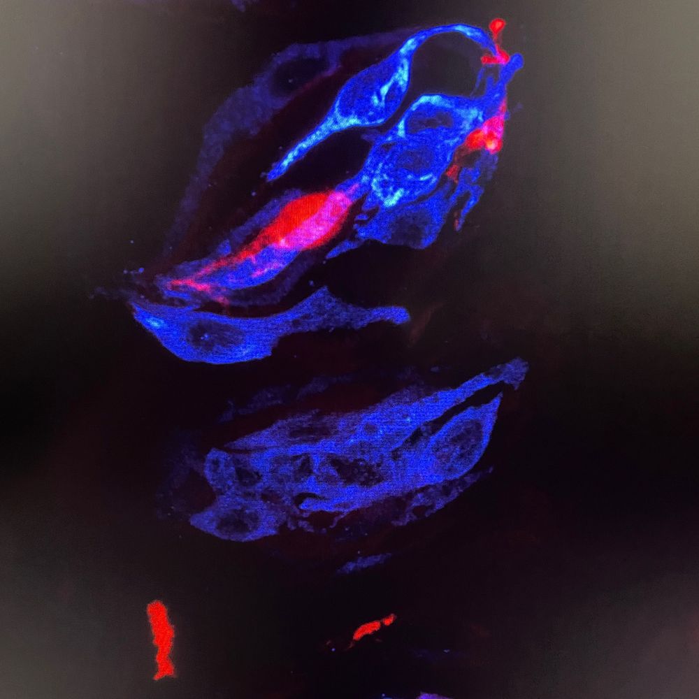
Tales from the Crypts!
... loading 85%
Exciting news coming soon!
#FluorescenceFriday
#devbio
pic credit: Ulrik Larsen
16.01.2026 18:34 — 👍 10 🔁 1 💬 0 📌 0
Love Berlin, but Norway beats it anytime 😉
16.01.2026 16:20 — 👍 0 🔁 0 💬 1 📌 0
1. Apply for one of the positions below (lab or theory) by Jan 18th
2. Learn interdisc skills and discover cool new things about how mitochondria move and socialise
3. Explore some of Norway's beautiful nature (both the below, Rundemanen and Gullfjellet, <10km from work)
09.01.2026 08:02 — 👍 5 🔁 8 💬 0 📌 0

New year - new socials !
Finally found a bit of time to make the transition from X to a more serene sky. Happy to be here 🙂
Below a picture from one of our recent studies showing lipid droplets staining in mouse liver upon high fat diet:
14.01.2026 00:14 — 👍 7 🔁 0 💬 0 📌 1