High-resolution in vivo kinematic tracking with customized injectable fluorescent nanoparticles
Injectable fluorescent nanoparticles were used to track positions on and inside of freely moving animals at high resolution.
Our first preprint has been accepted for publication www.science.org/doi/10.1126/... !!
tldr: @ezeyulu00.bsky.social , @amartyapradhan.bsky.social , @dkoveal.bsky.social and I developed a method using injectable nanoparticles to turn mice into…constellations in motion. 🧵⤵️
02.10.2025 19:45 — 👍 33 🔁 10 💬 2 📌 4
This work was driven by the singular vision and dedication of Zeynep Ulutas, @ezeyulu00.bsky.social . We could not have finished this without @amartyapradhan.bsky.social . @dkoveal.bsky.social provided key guidance on chemistry.
02.10.2025 19:45 — 👍 2 🔁 1 💬 0 📌 0

a man in a suit and tie is dancing in a room in front of a wall .
ALT: a man in a suit and tie is dancing in a room in front of a wall .
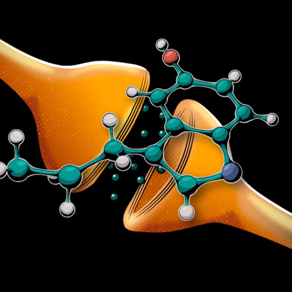
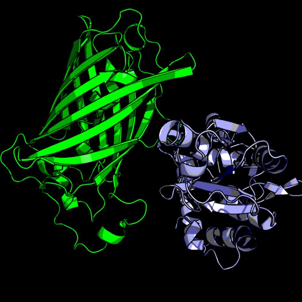
Beautiful structural analysis of a lifetime sensor from Rosen et al. Really cool mechanism reveal - something I never would have guessed when we made LiLac. A must-read for sensor folks!
www.pnas.org/doi/10.1073/...
09.03.2025 19:33 — 👍 2 🔁 1 💬 0 📌 0
Could one envision a synthetic receptor technology that is fully programmable, able to detect diverse extracellular antigens – both soluble and cell-attached – and convert that recognition into a wide range of intracellular responses, from gene expression and real-time fluorescence to modulation..
04.12.2024 16:05 — 👍 434 🔁 138 💬 33 📌 17

Figure 1. Screening and characterization of glucose sensors.
A) The screening assay identifies responsive glucose sensors in a medium-throughput manner. Plates lacking glucose have YSH757 yeast cells expressing sensor variants and are visualized whereafter glucose is sprayed on the plate and visualized again. Dividing the 2 images yields a ratiometric image showing which colonies have a responsive sensor. Colour indicates ratio of fluorescence after the spray divided by the fluorescence before the spray. B) normalized change of each colony tested on the plate, colour indicates basal fluorescence. C) design of the best performing variant. D) In-vivo CFP emission spectrum of YSH757 cells expressing TINGL. Colour indicates amount of glucose added to the cells. Data obtained in a fluorescence plate reader E) In-vivo Dose-response curve of YSH757 cells expressing TINGL or TINGL-RR obtained using a plate-reader. Fluorescence and CFP:RFP ratio are in absolute values. Line shows a Hillfit through the data points. F) Dose-response curve of TINGL-RR of cell-free extracts. Top facet shows CFP:RFP ratio versus mM glucose with the shapes depicting the pH. Line shows a Hillfit through the data of pH 7.5, 7 and 6.5 combined. Colored panel indicates the physiological pH of yeast.
Great to see this online as #preprint:
TINGL - A Turquoise INdicator for GLucose by the lab of @basteusink.bsky.social (and a small contribution from me): www.biorxiv.org/content/10.1...
03.12.2024 21:41 — 👍 28 🔁 6 💬 0 📌 0
Yulong Li lab at Peking University
Rural neuroscientist. Connectomics. Protists.
caveat emptor: https://bsky.app/profile/dddavi.bsky.social/post/3k5bnogiq5r2j
Husband, Father, Grandfather, Datahound, Dog lover, Fan of Celtic music, Former NIGMS director, Former EiC of Science, Stand Up for Science advisor, Shenanigator, Pittsburgh, PA
NIH Dashboard: https://jeremymberg.github.io/jeremyberg.github.io/index.html
Chair, Dept. Cell Biology at Emory SOM, Atlanta
RNA and synapse biology in neuronal health and disease
https://cellbio.emory.edu/basselllab/
https://med.emory.edu/departments/cell-biology/index.html
Neuroscience lab, neuromodulation, optogenetics, tool development, serotonin, neuronal circuits
Glasshalfemptologist.
https://drugmonkey.wordpress.com/
I talk about computational #openscience communication and science publishing. On the jupyterbook.org & @mystmd.org teams, co-founder of @curvenote.com & @continuous.foundation.
Neuroscience 🧠 PhD Candidate at #EmoryUniversity and #GeorgiaTech studying #psychedelics’ effect on 🐁 behavior | Previously at the Tata Institute of Fundamental Research
Neuroscience PhD student at Harvard
章雪翎 she/her from Guangzhou
🧠working at #MarkoLab
Smith College 2022
Neuroscience PhD Candidate
#EmoryUniversity and #GeorgiaTech
🇸🇪 Sweden's national center for molecular biosciences
👩🔬 Infrastructure with unique technologies and expertise
👥 Research community
🥼 Life science 🌱
💻 Often data-driven
https://www.scilifelab.se/
https://www.linkedin.com/company/scilifelab/
Nature Portfolio’s high-quality products and services across the life, physical, chemical and applied sciences is dedicated to serving the scientific community.
NeuroCyto lab 🤹🏻 Institute of NeuroPhysiopathology at CNRS & Aix-Marseille Université 🧠 Neuronal cell biology 🦠 Super-resolution microscopy 🔬 Come for science 👨🏻🔬 Stay for some cooking, sneakers and bike adventures 🥘 👟🚴
Professor at Karolinska Institutet specializing in the development and application of advanced cell imaging techniques to uncover and understand cell signaling mechanisms in health and disease.
Assistant Professor at Division of Molecular Neurobiology, Karolinska Institutet, Stockholm, Sweden.
Neuroscience | Neuroplasticity | Extrinsically Ordered/Disordered Proteins
Group page:
ki.se/en/research/groups/dagliyan-lab
I make fluorescent indicators that monitor metabolites and signaling molecules in the cell.
Now @KULeuven, previously in @DKFZ and @VIBLifeSciences.
Dedicated to drug discovery, enthralled by science. 5 kids. She. SAB Recursion. Founder SyzOnc. Lab at Broad Institute. Opinions my own.