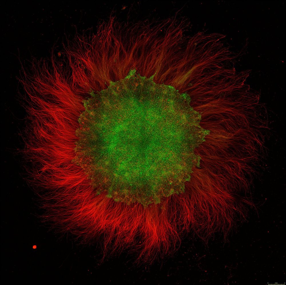
"A Sparkling Spot" ✨
🔬: Leica Biosystems SP8 Confocal, White Light Laser
🥼: Keunjung Heo, FM Kirby Neurobiology Center
📝: Harvard Brain Science Initiative Beauty of the Brain Awards: mailchi.mp/brain.harvar...
🏛️: Boston Children's Hospital (CIC): research.childrenshospital.org/resources/co...
09.02.2026 15:52 — 👍 0 🔁 0 💬 0 📌 0
📢 Calling the #NESM community!
We’re coming up on a year of sharing incredible microscopy data — and we want you to be part of it 🔬✨ Submit your work and we’ll feature it in future posts.
📩 Submit here: forms.gle/5bApb3RTVoA1...
#Microscopy #ScienceCommunication #Imaging
09.02.2026 15:51 — 👍 2 🔁 0 💬 1 📌 1

For #MicroscopyMonday, meet Rylie Walsh — Imaging Specialist at @mblscience.bsky.social and longtime board member. Our Director of Biological Sciences, she is passionate about helping scientists design #microscopy experiments, and is excited by all things #VolumeEM! 🔬
@ryliewalsh.bsky.social #NESM
02.02.2026 15:23 — 👍 3 🔁 0 💬 0 📌 0


For #MicroscopyMonday, meet our #NESM President, Jaqulin! She studies how alpha-synuclein affects synapses in sea lamprey at @mblscience.bsky.social, and is passionate about microscopy education & outreach.
🔬: @jeolusa.bsky.social 120i TEM
🐍: Sea lamprey reticulospinal synapse
🥼: Jaqulin Wallace
26.01.2026 14:57 — 👍 3 🔁 0 💬 0 📌 0
The Allen Mouse Brain Atlas is a cornerstone of #neuroscience: a standardized, high-resolution map of brain anatomy built to keep experiments comparable across labs. 🧠 Here’s a 3D walk through its anatomical meshes in #syGlass!
@alleninstitute.org
#3DThursday #DataViz #VR
22.01.2026 15:29 — 👍 10 🔁 6 💬 0 📌 1

For today’s #MicroscopyMonday, meet our #NESM President-elect, Harry Cramer! The Assistant Director of Cellular Imaging Core at Boston Children’s Hospital, Harry brings deep expertise in multiphoton microscopy, training, and community-driven science. 🔬🧠
@bostonchildrens.bsky.social
12.01.2026 15:11 — 👍 1 🔁 0 💬 0 📌 0


Introducing #NESM Treasurer Jennifer Misuraca for #MicroscopyMonday! Jennifer is FESEM Product Manager at #JEOL USA, supporting applications, training, and demos. She joined the NESM board in 2016 and became Treasurer in 2024. 📸 SEM of bacteriophage on a JEOL FESEM, Joseph Stukey at Hope College
05.01.2026 17:47 — 👍 2 🔁 0 💬 1 📌 0
Happy Holidays from NESM!
25.12.2025 17:08 — 👍 1 🔁 0 💬 0 📌 0


For #MicroscopyMonday, we’re excited to welcome Katie Shulenberger as NESM’s newest Director of Physical Sciences! 🎉
An Asst. Prof. of Chemistry at @brandeis.edu, Katie develops spectroscopic methods to study semiconductor nanomaterials and photophysics.
#NESM #Spectroscopy #Nanomaterials
22.12.2025 16:48 — 👍 4 🔁 0 💬 0 📌 0


Another #MicroscopyMonday intro! Meet Emma Perry, the #NESM Clerk from UMaine @umaine.bsky.social . She manages electron & confocal microscopes at UMaine and studies Maine/NE USA tardigrade biogeography & taxonomy. 📸 Maine tardigrade: Viridiscus perviridis 🔬Amray 1820, CORE #ElectronMicroscopy Lab.
15.12.2025 14:58 — 👍 2 🔁 0 💬 0 📌 0
The New England Society for Microscopy is heartbroken by the tragic shooting at Brown University. Academic and research spaces should be safe places to learn, teach, and experiment. We mourn the victims, stand with the Brown community, and are deeply angered by this senseless act of violence.
14.12.2025 15:38 — 👍 3 🔁 0 💬 0 📌 0


Introducing another #NESM board member for #MicroscopyMonday — Ben Muller, Director of Physical Sciences! He works across surface metrology, microfluidics, thin films, and micro/nanofabrication. He just defended his PhD! 🎉
📸 SEM of modified EGaIn sphere contacts (Harvard CNS).
#SEM #Nanotechnology
08.12.2025 13:28 — 👍 2 🔁 0 💬 0 📌 0
#NESM team together!
04.12.2025 17:59 — 👍 1 🔁 0 💬 0 📌 0

Happy #MicroscopyMonday! Meet our new Director of Biological Sciences, Mark Scimone, a UNH professor whose work spans neuroscience, optics, and image analysis. He brings experience from Boston Children’s and PSI, plus a passion for microscopy education. #NESM #SEM
📸: doi.org/10.1038/s415...
01.12.2025 15:27 — 👍 5 🔁 0 💬 0 📌 0
Happy #MicroscopyMonday! This week we're spotlighting Brandy Freitas, our Director of Media and Outreach at #NESM.👏 @brandy-syglass.bsky.social
Brandy is the Director of Data Science at #syGlass, and here is a video of #3Ddata from C Wolff the MBL @mblscience.bsky.social 🔬
#BioImaging #SciComm
23.11.2025 19:50 — 👍 6 🔁 0 💬 1 📌 1

Happy #MicroscopyMonday! 🧫 This week we're spotlighting Andy Stone, our immediate past-president at #NESM. 👏 @andystoneimaging.bsky.social
Andy leads the Brandeis light microscopy core and is a powerhouse in the imaging community—from BINA to NESM and RMS. 🎥🔬
#LightSheet #BioImaging #STEM
17.11.2025 17:16 — 👍 10 🔁 2 💬 0 📌 0
Great!
13.11.2025 13:47 — 👍 1 🔁 0 💬 0 📌 0
This gorgeous piece will be displayed during our Fall Symposium at Brandeis University in Waltham, MA on November 6th & 7th. If you are interested in a chance to learn more about 3D printing and how it can be used in your microscopy builds, register now for the Makerspace Workshop on November 6th.
27.10.2025 14:59 — 👍 0 🔁 0 💬 0 📌 0
This week's #MicroscopyMonday is a time lapse 3D printing of a mini microscope from the MakerLab at Brandeis University.
🥼 Greg Buckland | Head of Brandeis MakerLab
#MicroscopyMonday #Sciencecommunication #BrandeisUniversity #NESM #Microscopy #Science #3D #3DPrinting
27.10.2025 14:59 — 👍 7 🔁 0 💬 1 📌 0

We're up in Gloucester today for the 10th Annual GMGI Science Forum! #NESM #GMGI
23.10.2025 13:35 — 👍 2 🔁 0 💬 0 📌 0

Tungsten crystals for #MicroscopyMonday from one of our upcoming keynote speakers! #NESM www.nesmicroscopy.org/2025-fall-sy...
🥼: @gewilli.bsky.social, Microscopist
🏛️: Brown University Leduc Bioimaging
🔬: TFS Apreo 2 VS Scanning Electron Microscope, Horizontal Field Width (HFW) = 49μm
20.10.2025 16:29 — 👍 2 🔁 0 💬 0 📌 0

It's #MicroscopyMonday and we are only three weeks away from the #NESM Fall Symposium! We still have opportunities for poster and oral presentations for researchers, vendors and core facilities, link here: www.nesmicroscopy.org/2025-fall-sy...
#Microscopy #LightSheet #3DPrinting #SEM #LSFM #SciComm
13.10.2025 16:30 — 👍 2 🔁 0 💬 0 📌 0

For #MicroscopyMonday: “Seeing Every Photon”—a hands-on workshop with Evident Scientific on the FLUOVIEW FV4000 confocal! At the Fall symposium Nov 6-7, learn to capture consistent, quantitative images with SilVIR™ detectors and performance monitoring. #NESM #ConfocalMicroscopy
06.10.2025 11:55 — 👍 2 🔁 0 💬 0 📌 0

Stylized graphic of an electron beam, shown as a purple cone with a red bullseye tip, aimed precisely at a single atom in a hexagonal lattice of atoms. The background is a faint atomic-resolution microscopy image, highlighting the idea of targeting individual atoms in atomically thin materials
Excited to welcome MIT’s Julian P. Klein as a keynote at the #NESM Fall Symposium! 🔬 His work uses electron beams to target atoms in 2D materials — here’s a teaser image from his research in quantum engineering.
#MicroscopyMonday #ElectronMicroscopy #QuantumMaterials #Nanotechnology #MIT #SciComm
29.09.2025 15:43 — 👍 2 🔁 0 💬 1 📌 0

📣Announcing the Fall Symposium Workshops for #MicroscopyMonday!🎫 Registration is now open here: www.nesmicroscopy.org/2025-fall-sy...
🔬: Hitachi Gemini FE SEM
🥼: Benjamin Muller, PI Giovanni Traverso, MIT Department of Mechanical Engineering (MechE)
📝: Feig et al, lnkd.in/ea5M7aET
#NESM #SciComm
22.09.2025 16:01 — 👍 1 🔁 0 💬 0 📌 0
Announcing one of our Keynote Speakers, Elizabeth Engle, for #MicroscopyMonday! Please join us in November at Brandeis!
🔬: 3i Light Sheet
🐭: Dharmendra Puri, Boston Children's Hospital
🏛️: Harvard Medical School's MicRoN facility
🎬: syGlass.io
#NESM #LightSheetMicroscopy #3DImaging #TissueClearing
15.09.2025 16:50 — 👍 8 🔁 1 💬 0 📌 0

Announcing registration for the Fall Symposium at Brandeis for #MicroscopyMonday! #NESM
www.nesmicroscopy.org/2025-fall-sy...
🔬: Bruker TruLive3D @brukerspatial.bsky.social
🥼: Amro Hamdoun @hamdounlab.bsky.social , Tzer Han Tan's @tzerhan29.bsky.social group
🌌: Painted Sea Urchin
📷: syGlass.io
08.09.2025 14:13 — 👍 4 🔁 3 💬 0 📌 0

🕷️ A winter tick’s lethal grasp for a late #MicroscopyMonday, captured in stunning SEM by Monica Miles, winner of 3rd place in UMaine’s 2025 Imaging Your Research Contest.
Tiny structures, huge impact on Maine’s moose calves.
#Microscopy #Ecology #Moose #SEM #NESM @umaine.bsky.social
02.09.2025 15:25 — 👍 2 🔁 0 💬 0 📌 0
It's #MicroscopyMonday! Today we have a very special confocal dataset that HIGH SCHOOL students were able to label and image themselves at the MBL.
🐟: #Zebrafish
🔬: Nipam Patel, High School Science Discovery Programs
🏛️: @mblscience.bsky.social
🎬: syGlass.io
#NESM #Confocal #SciComm #ScienceEd
25.08.2025 20:32 — 👍 6 🔁 1 💬 0 📌 1
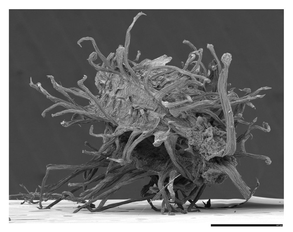
Scanning Electron Microscope image of a Daucus carota seed pod.
Black line on the bottom of 667 μm long for scale.
Dedicated seed pod of Wild Carrot/Queen Anne's Lace/Daucus carota
📷 (Ok maybe 🔬) Hitachi 2700 SEM
More of the #SciArt side.
(I'll be giving a talk about it all in November at the @nesmicroscopy.bsky.social fall meeting, not this plant as much but related)
20.08.2025 15:15 — 👍 19 🔁 2 💬 0 📌 0
International network of young scientists sharing imaging and analysis challenges in a friendly, peer-based space. We offer flexible learning via watch parties, roundtables, troubleshooting sessions and expert-led events.
https://gerbi-gmb.de/teams/ymia/
Relaying microbiology news, articles and comments relevant to aspects on bacteria, fungi, viruses and other microbes / microorganisms. Microbes are 💪
New submissions in arXiv
Category: Condensed Matter
https://arxiv.org/list/cond-mat/new
Administrator: krypf
@krypf.bsky.social
lit link: lit.link/krypf
I host that Ologies podcast
A comedyish science podcast hosted by @alieward. Asking smart ologists not-smart questions about their professional obsessions. Every Tuesday. May be NSFW. https://linktr.ee/alieward
Ph.D. ex-academic, self-published author of “A Heuristic Guide To Quantitative Imaging”. Lead, operations and technical sales at Coastal Microscopes.
Bioimage analysis and computer vision enthusiast. Whenever I can, I do software too.
Ikerbasque | University of the Basque Country (UPV/EHU) | DIPC | Biofisika Institute
ORCID: 0000-0003-0229-5722
A complete resource for the latest discoveries in cell biology, from organoids to immunology. Brought to you by STEMCELL Technologies.
Digital image analyst @mdibiolab.bsky.social @mdibl-lmf.bsky.social
#science #microscopy #mdibiolab #bluesci
The Centre of Marine Sciences (CCMAR) is a non-profit organization located on the Gambelas campus of the University of Algarve and dedicated to R&D in the Marine Sciences.
Quantitative Imaging: From Acquisition to Analysis course at Cold Spring Harbor Laboratory / Posts by QI Instructors & TAs
Applications for QI 2026 are due 1/30!
https://meetings.cshl.edu/courses.aspx?course=C-QICM
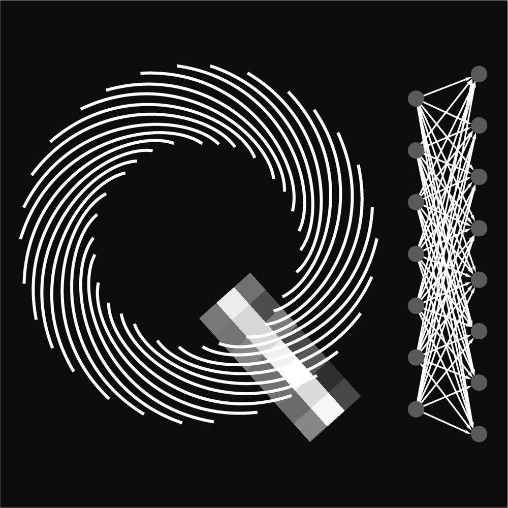
Physicist by training, working in computational neuroscience at the @dzne.science. Interested in how neural dynamics and learning shape brain function and behavior, using modeling and data analysis.
🌍fabriziomusacchio.com
🐘mastodon.social/@FabMusacchio
FocalPlane is a community site for anyone with an interest in microscopy. Hosted by Journal of Cell Science (@jcellsci.bsky.social) and The Company of Biologists (@biologists.bsky.social).
https://focalplane.biologists.com/
Itinerant cycligrapher©™
Coffee, Bikes, Photography, Food, you know that stuff
Brain scientist who studies how we hear and form memories of sounds 🧠 🧬🔬🧪; prof at Penn. Loves🧘♀️🚴♀️✈️🥐🧶📷⛷️🏔️. Mom to 👦👧👦🐈🐈. Lab: hosting.med.UPenn.edu/hearing
Cell and Developmental Biologist @ St. Jude Children's Research Hospital - 🦔 signaling in 🧠🫀&🫁 - science 🧪, academia, DevBio research, mentoring, scicomm, 🔬, birds 🪶, hikes - fueled by ☕ & 🧘🏼♀️ - Y’all means all - Views my own and not SJCRH's
PhD in the lab of @vlecaudey.bsky.social at JGU. Developmental Biology in #Zebrafish.
PharmD, MS in Biophysics, PhD in Biochemistry
I like taking photos of weird things inside the brain 🔬
Cure #LongCovid
Long Covid Advisory Team: https://whn.global/long-covid-advisor.
daniellebeckman.com