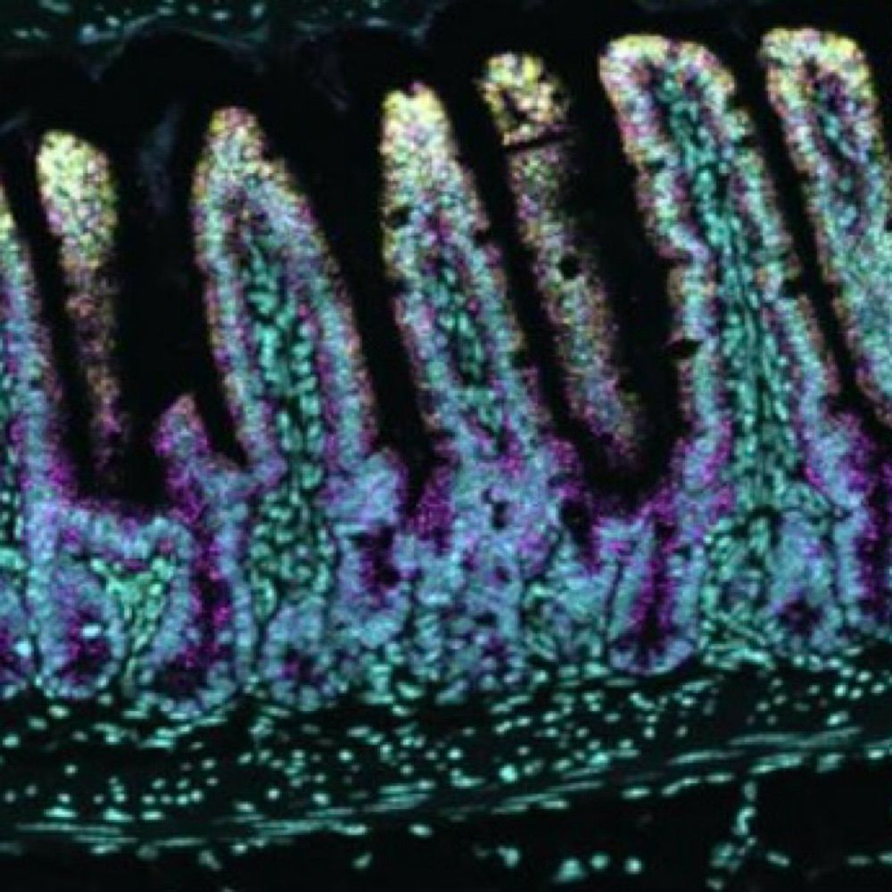
#MicroscopyMonday featuring gastric metaplasia. Extracellular matrix staining is purple, enteroendocrine cells are yellow, and mucus cells are teal.
03.02.2026 03:34 — 👍 5 🔁 2 💬 0 📌 0
@amyengevik.bsky.social
Epithelial cell biologist and physiologist studying the GI tract.
#MicroscopyMonday featuring gastric metaplasia. Extracellular matrix staining is purple, enteroendocrine cells are yellow, and mucus cells are teal.
03.02.2026 03:34 — 👍 5 🔁 2 💬 0 📌 0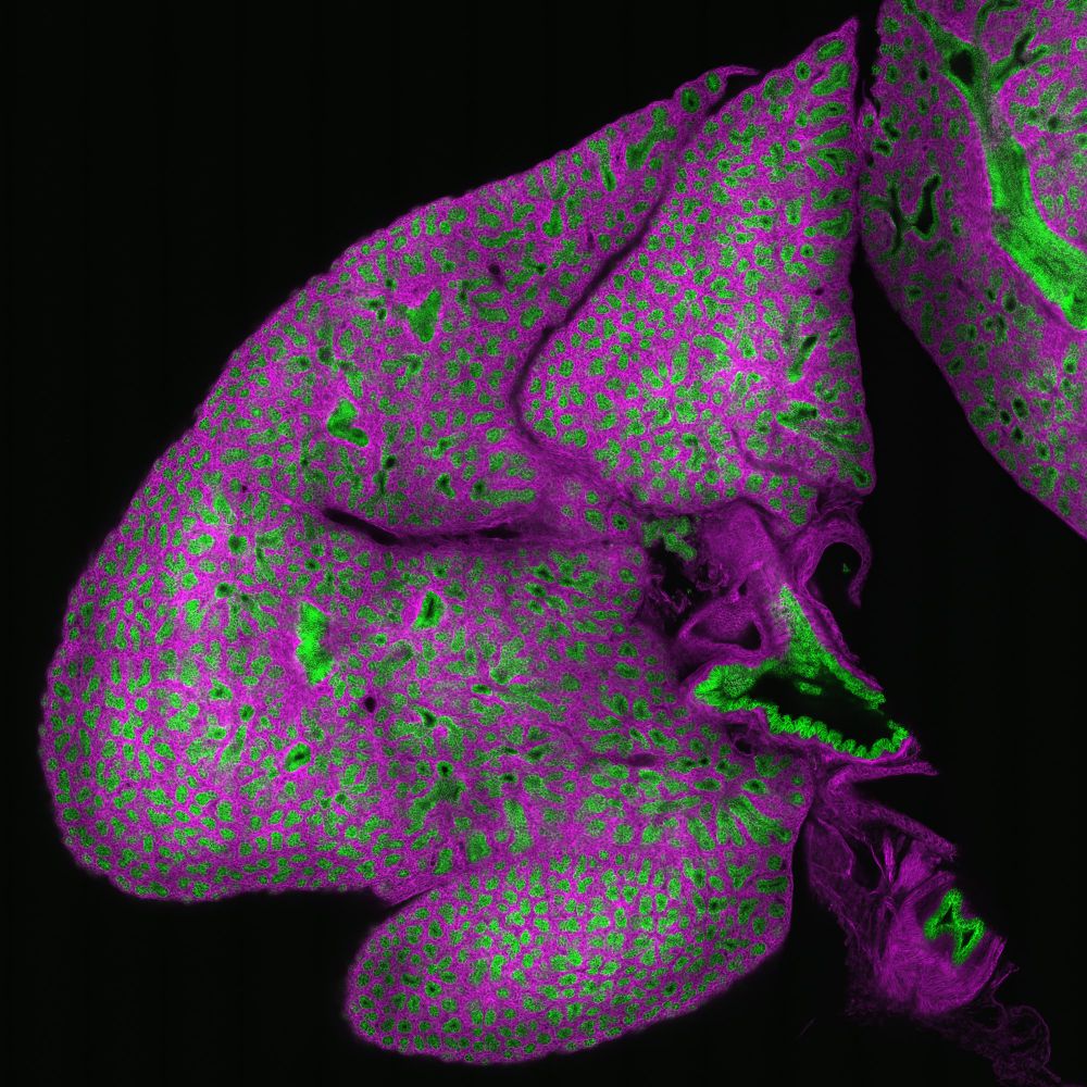
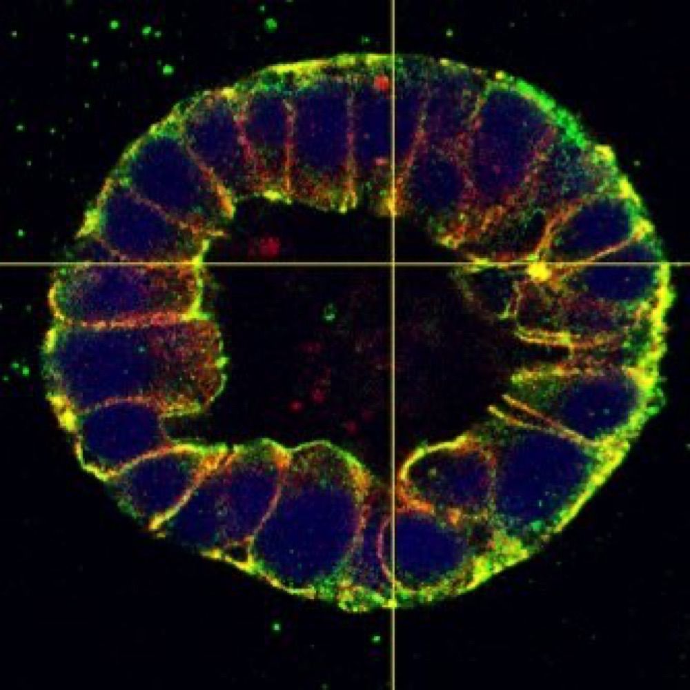
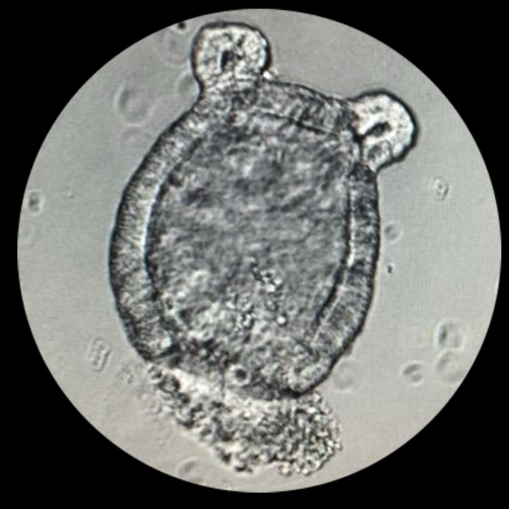
Microscopy image of a developing mouse lung. Cells that express Shh are shown in green. Membranes of non-expressing cells are magenta.
Happy #FluorescenceFriday! I've shared this before but am reposting b/c I think the developing #lung is just so darn pretty! This is an E18.5 🐭 lung with 🟢 marking cells that express my favorite gene, Shh.🟣 marks cell membranes. 🔬 by @stjuderesearch.bsky.social postdoc👩🔬 @christinaadaly.bsky.social 🧪
23.01.2026 14:35 — 👍 37 🔁 8 💬 0 📌 0
A confocal microscopy image showing early tau accumulation in the brain with Alzheimer's Disease. Everything in blue (all nuclei) and green (neurons) is healthy. All the other colors are composed of a combination of different markers for pathological tau. The key question is: how do we avoid this tau spreading from cell to cell via neuronal connections? If we can avoid this, we can avoid memory loss.
Guess you can use a timeline cleanser? Can I show you some microscopy? My job is to see beauty even in sad things (so we can learn how to prevent them!). This is a brain with early Alzheimer's. This image was acquired in 3D and took 72 hours to get ready!
#Microscopy #Neuroscience

Different small intestinal cell types for #FluorescenceFriday
31.01.2026 03:50 — 👍 24 🔁 1 💬 0 📌 1
#TuftTuesday featuring some interesting tuft cell (pink) morphologies.
28.01.2026 03:04 — 👍 6 🔁 0 💬 0 📌 0
#MicroscopyMonday
26.01.2026 22:18 — 👍 6 🔁 0 💬 0 📌 0
Happy #FluorescenceFriday from some small intestinal villi.
23.01.2026 22:56 — 👍 11 🔁 1 💬 0 📌 0
So many tuft cells (yellow/orange) for #TuftTuesday. Immune cells are in green.
20.01.2026 22:03 — 👍 15 🔁 1 💬 1 📌 0
Some intestinal mucus staining (green) for #MicroscopyMonday
20.01.2026 04:12 — 👍 11 🔁 1 💬 1 📌 0
Happy #FluorescenceFriday!
17.01.2026 04:39 — 👍 30 🔁 4 💬 0 📌 0Thanks! My lab does our own sectioning. It saves a ton of money and gives us some control over our samples.
15.01.2026 01:40 — 👍 1 🔁 0 💬 0 📌 0
The first gland at the stomach’s limiting ridge is packed with tuft cells #TuftTuesday
14.01.2026 00:47 — 👍 5 🔁 0 💬 1 📌 0
A modified H&E for #MicroscopyMonday
13.01.2026 04:26 — 👍 9 🔁 0 💬 0 📌 0
The first image I took of 2026 #FluorescentFriday
10.01.2026 04:42 — 👍 36 🔁 2 💬 0 📌 0
First #MicroscopyMonday of 2026 deserves some festive cells.
06.01.2026 03:26 — 👍 18 🔁 5 💬 0 📌 0
Hoping everyone's new year fluorescence is clear and bright. Happy #FluorescenceFriday.
03.01.2026 04:52 — 👍 31 🔁 6 💬 0 📌 0
#GESRC GI Epithelium Meeting is wrapping up today. A great conference full of great research.
10.09.2025 21:07 — 👍 2 🔁 0 💬 0 📌 0
Enjoying #GESRC, so many good talks and a lot of interesting research on GI tuft cells #TuftTuesday
09.09.2025 23:25 — 👍 12 🔁 2 💬 0 📌 0Great question, it could use some neon tiger print to make it more Lisa Frank 🙂
20.08.2025 02:54 — 👍 1 🔁 0 💬 0 📌 0
Some tuft cells for #TuftTuesday
20.08.2025 02:53 — 👍 6 🔁 0 💬 0 📌 0
Is this too Lisa Frank for a journal cover? #MicroscopyMonday
19.08.2025 01:32 — 👍 56 🔁 5 💬 4 📌 0That’s such a good hashtag!
17.07.2025 21:48 — 👍 1 🔁 0 💬 0 📌 0
#TuftTuesday
01.07.2025 19:27 — 👍 11 🔁 0 💬 0 📌 0Thanks! It’s just one set of antibodies. I can add up to four plus DAPI.
01.07.2025 16:16 — 👍 1 🔁 0 💬 0 📌 0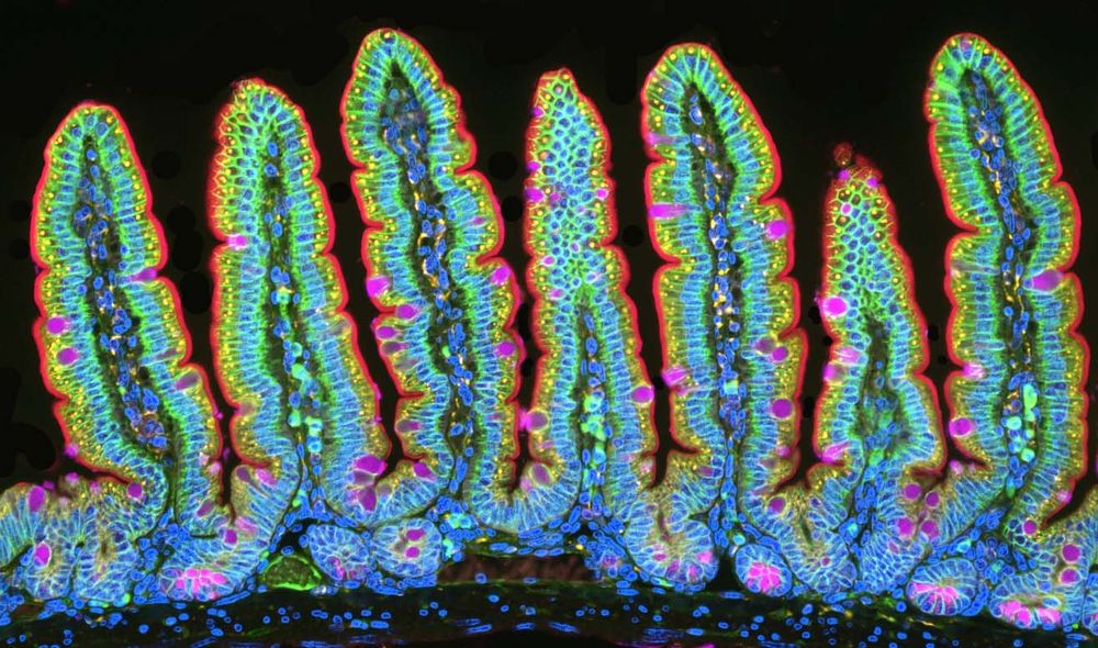
Goodbye June #MicroscopyMonday
30.06.2025 19:18 — 👍 34 🔁 4 💬 1 📌 0
Every week feels a bit longer, glad that it is finally #FluorescenceFriday. Mouse liver tissue featuring a lot of diploid cells.
27.06.2025 17:35 — 👍 24 🔁 2 💬 0 📌 0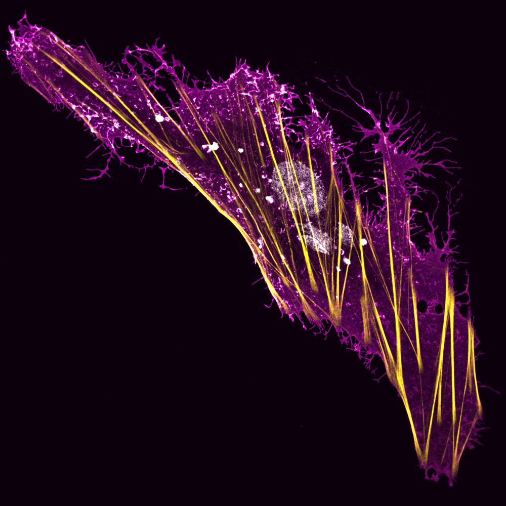
A Boa Constrictor brain-derived cell expressing a viral glycoprotein (magenta) and stained for actin (yellow) and DNA (white).
#FluorescenceFriday

Another #MicroscopyMonday featuring small intestinal cells
10.06.2025 03:55 — 👍 11 🔁 0 💬 0 📌 0
Happy Birthday! Wishing you all the best.
07.06.2025 02:41 — 👍 3 🔁 0 💬 0 📌 0
Liver immunostaining for this #FluorescenceFriday
06.06.2025 21:15 — 👍 29 🔁 5 💬 0 📌 0