In December's #EditorsChoice, Nayeli Reyes-Nava, @edwardmarcotte.bsky.social, @jbwallingford.bsky.social & co use #Xenopus model to study #Cilia motility disturbances in primary ciliary dyskinesia #PCD
⚡ doi.org/10.1242/dmm....
📰 doi.org/10.1242/dmm....
30.12.2025 12:01 — 👍 5 🔁 5 💬 0 📌 0
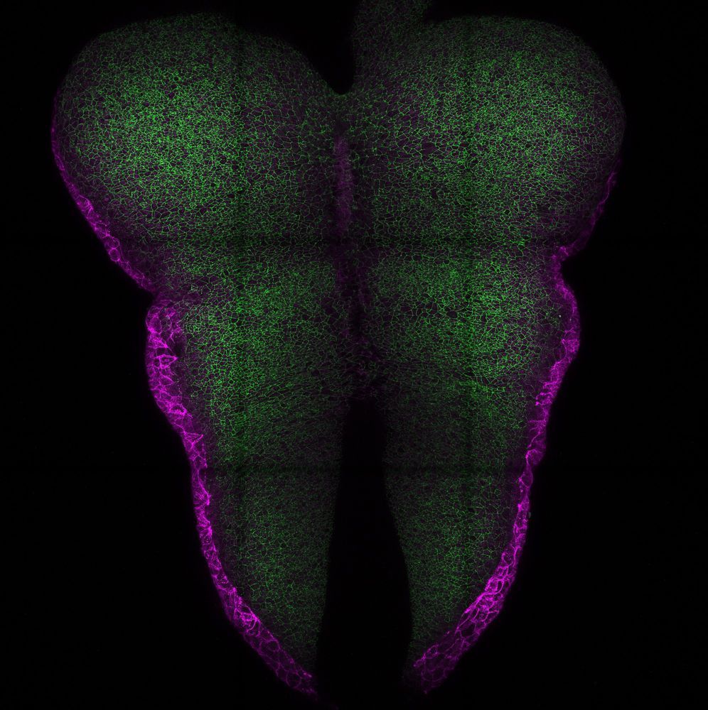
Genetically engineered ESC-derived embryos reveal Vinculin-dependent force responses required for mammalian neural tube closure https://www.biorxiv.org/content/10.64898/2025.12.22.696028v1
25.12.2025 18:30 — 👍 4 🔁 6 💬 0 📌 0
This was such a fun project where we uncovered many fascinating details, including a surprising regulatory layer to Hh signaling acting along the AP axis to influence both cell fates and the apical constriction program driving closure. Really grateful to Sun-Hee, Saikat and Kevin!
23.12.2025 16:20 — 👍 0 🔁 0 💬 0 📌 0

GPR161–GLI3 repressor signaling at cilia directs apical constriction and cell fate during cranial neural tube closure
Highlighted Article: Cilia-dependent GLI3 repression controls patterns of cell fate and apical remodeling during cranial neural tube closure in mouse.
The final manuscript detailing a great collaborative effort between Saikat Mukhopadhyay's group and ours, spearheaded by the amazing Sun-Hee Hwang is out now! We disambiguate the roles of activating and repressive arms of Hh signaling during cranial neural tube closure. doi.org/10.1242/dev....
23.12.2025 16:20 — 👍 2 🔁 0 💬 1 📌 0

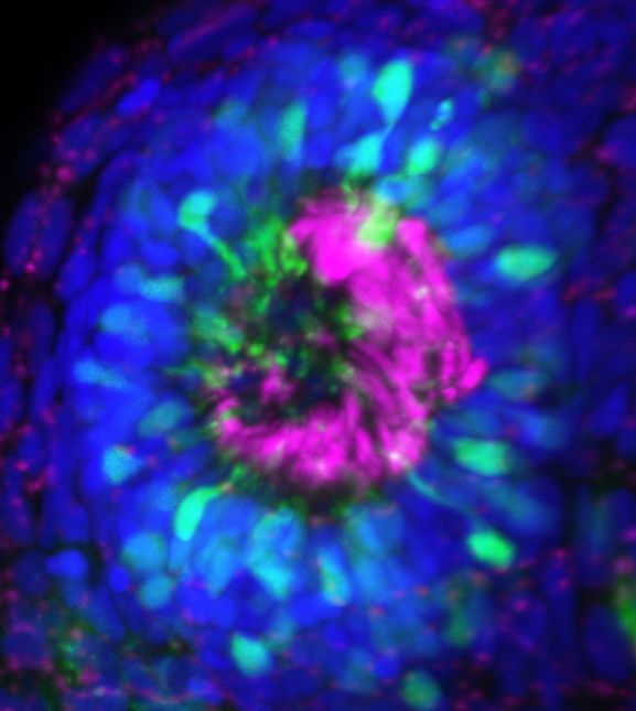
Floor plate marker ventralization in E9.25 Gpr161 ko forebrain is selectively rescued by Gli3R expression but not from Gli2 deletion. (A) Forebrain (top) and hindbrain (bottom) cranial horizontal sections immunostained using designated markers from embryos dissected at E9.25 (18-22 somites, except Gli2 ko/ko at 25 somites) of the following genotypes: wild-type (n=6), Gpr161ko/ko (n=3), Gli3RΔ701/+ (n=6), Gpr161ko/ko; Gli3RΔ701/+ (n=3), Gli2ko/ko (n=6), Gpr161ko/ko; Gli2ko/ko (n=6). The same sections for each genotype were co-stained for FOXA2 and NKX6-1 and consecutive sections for PAX6. All images are counterstained with DAPI. Scale bars: 100 µm. FB, forebrain; HB, hindbrain; OC, optic cup. See also Figs S2 and S3, which show magnified forebrain and hindbrain images of the same sections (except for staining of separate closed hindbrain sections for Gpr161ko/ko; Gli3RΔ701/+ and Gpr161ko/ko; Gli2ko/ko and additional staining for OLIG2 for all genotypes). Additional anterior to posterior sections in Gpr161ko/ko; Gli2ko/ko brain are shown in Fig. S2. (B,C) Quantification of FOXA2 dorsoventral (D-V) extent and ventral-most PAX6 extent with respect to full extent of the neural tube in forebrain and hindbrain regions. All data shown as mean±s.d. Note forebrain FOXA2 ventralization in Gpr161ko/ko rescued by Gli3R expression only, whereas hindbrain FOXA2 ventralization in Gpr161ko/ko is rescued by Gli3R expression or Gli2 deletion. Ventral-most PAX6 extents are not impacted from ventralized FOXA2 levels in Gpr161ko/ko; Gli2ko/ko. (D) Summary of the regionalized impacts on dorsoventral neural precursor patterning. WT, wild type.
The prickly consequences of Hedgehog de-repression on cranial neural tube closure
This Research Highlight showcases work by Eric R. Brooks @tomorrowbot.bsky.social, Sun-Hee Hwang, Kevin A. White and Saikat Mukhopadhyay:
journals.biologists.com/dev/article/...
23.12.2025 08:54 — 👍 4 🔁 2 💬 1 📌 0
Thanks to @devbiol.bsky.social for featuring our work! This was a great project, though one with many confusing turns at times, which only seems right for exploring something to do with such a re-iteratively used pathway as Wnt signaling
03.12.2025 15:23 — 👍 2 🔁 0 💬 0 📌 0
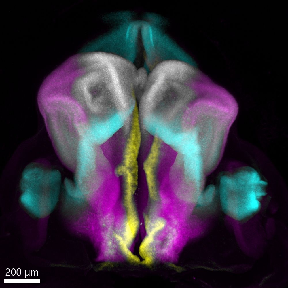
A look at the developing human embryonic forebrain in 3D!
Post-doc @stochastalive.bsky.social shares this week's image showing a dorsal view of the human embryonic forebrain at Carnegie Stage 16 (~6 weeks post conception). Major regions marked by FOXG1, WNT8B, PAX6, and SHH are displayed.
11.06.2025 16:54 — 👍 34 🔁 9 💬 1 📌 1
#ApicalConstriction and #DevBio afficionados - we've got two new pre-prints you may be interested in, below:
20.05.2025 14:30 — 👍 6 🔁 2 💬 1 📌 0
We are doing well and it's definitely neat to have a UT reunion here many years down the road!
07.05.2025 16:08 — 👍 1 🔁 0 💬 1 📌 0
Congrats on the big year, Ben!
06.05.2025 20:57 — 👍 1 🔁 0 💬 1 📌 0
WOW! Been a crazy busy year but we've gotten a lot of stuff across the finish line. All the credit to my team here at U of L and our collabs at IU SOM, Oxford, CU Anschutz and UC Merced! Gonna update y'all with this Bluetorial!
06.05.2025 17:57 — 👍 14 🔁 3 💬 3 📌 2
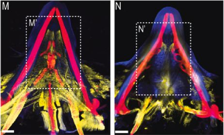
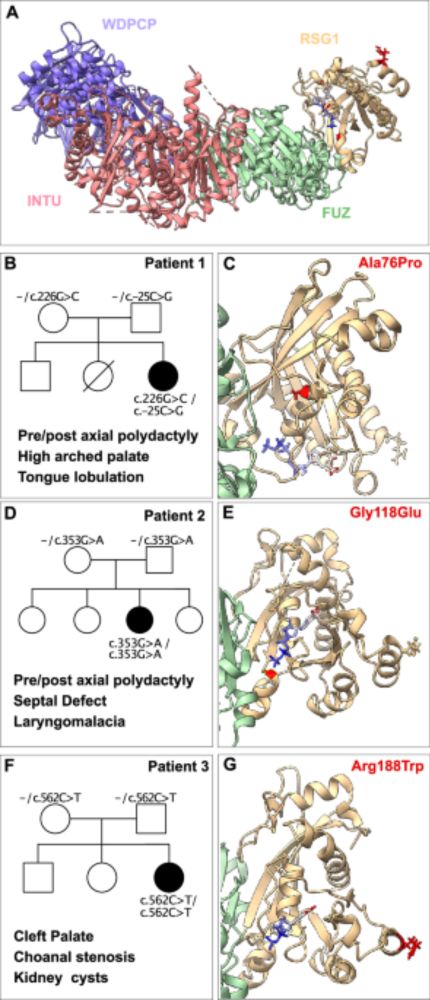
The ciliary protein C2cd3 is required for mandibular musculoskeletal tissue patterning. Evan C. Brooks, Ph.D. Graduate Student Alumnis, Brugmann Lab, Cinicinnati Children's Hospital Medical Center. Currently: Postdoctoral Fellow, University of Colorado School of Dental Medicine. "Biological Differentiation Across the Scales" Seminar, April 28, 2025.
The "Biological Differentiation Across the Scales" virtual seminar series kicked off on April 28 with an insightful, engaging talk by @ecbrooks96.bsky.social. Missed it live? Watch the recording: www.isdifferentiation.org/Journal/Page... @rogerslabucd.bsky.social @uribelab.bsky.social #devbio
06.05.2025 15:07 — 👍 4 🔁 7 💬 0 📌 0
On a final note, we would like to thank our reviewers for their enthusiasm about this work and for their experimental and analytical suggestions which significantly strengthened this study.
28.04.2025 17:23 — 👍 1 🔁 0 💬 0 📌 0
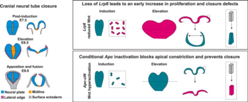
Thus, Wnt activity levels must be modulated throughout cranial neural tube closure in order to control both proliferation and cell remodeling in order for closure to complete.
28.04.2025 17:23 — 👍 1 🔁 0 💬 1 📌 0
Conversely, conditional inactivation of APC and resulting hyperactivation of Wnt signaling, leads to apical constriction blockade by altering actin organization, and this occurs without changes in proliferation or tissue scaling.
28.04.2025 17:23 — 👍 1 🔁 0 💬 1 📌 0
Loss of robust Wnt signaling in Lrp6 mutants drives an early burst of excessive cell proliferation in the cranial tissues, leading to a doubling of the tissue volume and width. This excessive width blocks closure despite the normal function of cell remodeling programs, including apical constriction.
28.04.2025 17:23 — 👍 1 🔁 0 💬 1 📌 0
In this study we find that either hypomorphic or hypermorphic Wnt signaling states lead to cranial neural tube closure defects, but interestingly the underlying cellular defects in pathway activity states are distinct.
28.04.2025 17:23 — 👍 1 🔁 0 💬 1 📌 0
Join us for the @socdevbio.bsky.social Southeast Regional Meeting, May 19-21! www.sdbonline.org/sesdb2025 Showcase your exciting research & receive valuable feedback at any stage, from a newly fertilized idea to a rapidly gastrulating hypothesis to a fully 'developed' story. 🙂 #SESDB2025 #DevBio
08.04.2025 23:46 — 👍 2 🔁 2 💬 0 📌 1
My daily haiku from grant writing:
More words fall beneath
The stoke of my delete key
Concision triumphs
17.01.2025 01:14 — 👍 1 🔁 0 💬 0 📌 0
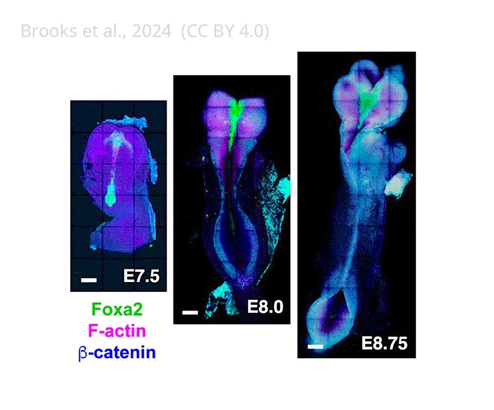
Images of mouse embryos spanning the stages of cranial neural tube closure
A new single-cell atlas of gene expression provides insights into the patterning of the neural plate of mice.
🔗 https://elifesciences.org/articles/105042?utm_source=bluesky&utm_medium=social&utm_campaign=organic_insights
07.01.2025 14:59 — 👍 5 🔁 4 💬 1 📌 0
Together, these analyses suggest that Wnt activity levels must be strictly modulated to tune both tissue scaling and curvature change during cranial closure.
20.12.2024 17:01 — 👍 0 🔁 0 💬 0 📌 0
On the other hand, hyperactivation of Wnt signaling by conditional inactivation of APC specifically in the cranial folds blocks apical constriction, preventing neural fold elevation.
20.12.2024 17:01 — 👍 0 🔁 0 💬 1 📌 0
We find that reduced Wnt signaling in Lrp6 mutants leads to a surprising tissue hyperplasia phenotype in the cranial neural plate, which leads to a doubling of tissue width, preventing closure. This hyperplasia is driven by an early doubling of proliferation specifically in anterior cranial tissues
20.12.2024 17:01 — 👍 0 🔁 0 💬 1 📌 0
Assistant Professor, Dept of Cell Biology, JHU SOM. Lipids and membranes in neural crest development. He/him. 🏳️🌈🐣🔬
https://piacentinolab.com/
Assistant Professor at University of Michigan. Developmental biologist using embryology, genetics, and imaging to discover how our organs form. She/her/hers. www.edwards-lab.org
Bioinformatics Scientist / Next Generation Sequencing, Single Cell and Spatial Biology, Next Generation Proteomics, Liquid Biopsy, SynBio, AI/ML in biotech // http://albertvilella.substack.com
Developmental Biology | Cilia | Scoliosis
Cellular and Molecular Biology PhD Candidate @UTAustin
Ryan Gray and John Wallingford Lab 🐟🐸
Engineering cells to understand their decisions
ESPOD Fellow @ebi.embl.org & @sangerinstitute.bsky.social
(Saez-Rodriguez & Parts)
lingering scientist @crick.ac.uk (Briscoe)
InSDB is the national society that represents all developmental and stem cell biologists in India
Embryo Enthusiast | Frog Fiend | Protein Person
🧬 PhD Candidate @ UT Austin
🐸💻 Wallingford and Marcotte Labs
🐅 Princeton University '20
Brain development postdoc in @CorinneHouart lab at KCL's
@dev_neuro and @TheCrick. Alumni of @WellcomeTrust PhD at @Cambridge_Uni and @uOttawa
Our lab studies fundamental mechanisms in early neurodevelopment and neurodegenerative diseases | Centre for Developmental Neurobiology @ King's College London | Satellite lab @ The Francis Crick Institute
https://ritolab.org | Human Development | Tissue Engineering | Stem Cell biology
Neuroembryologist. Mechanobiologist. Vet.
Developmental neurobiologist interested in evo-devo, neurogenesis, plasticity & neurodevelopmental disorders.
Professor @usc.gal
#NeuroDevo #EvoDevo #DevBio #sharks #retina
ICREA Research Professor. Evolution, Genetics, Neuroscience, Linguistic Cognition
https://www.cedricboeckx.com
Postdoc in Kyra Campbell's lab at the University of Sheffield - EMT, polarity and midgut morphology.
Board game fan (not that I ever win) and musician (to the annoyance of everyone around me) when I'm not at the microscope
Cardiac Development • Epigenetics • Postdoc • UPenn #devbio 🫀🔬🧬🐭 #firstgen #academicjobmarket
🚨Posts may contain science, bad jokes, cooking 🧵s, 🎨 &/or political snark. The typos are free.
Postdoc at the Vierbuchen lab (MSKCC), previously PhD at Institut Imagine (Cantagrel/Colleaux lab) /// I do mouse brain organoids to characterize early development and understand what makes each brain unique
Postdoc @ Sloan Kettering Institute interested in all things mechanobiology during cell and tissue morphogenesis. Past: PhD @Vanderbilt
Developmental Biologist working at the Francis Crick Institute. Neural tube, morphogens, and gene regulatory networks. Editor-in-chief, Development.
London · briscoelab.org
Bioscientist. Associate Professor at Rice University. Neural crest, zebrafish, enteric nervous system, gut-brain axis, neurodevelopment. Editor-in-Chief, Differentiation.